| RIKEN Center for
Developmental Biology (CDB) 2-2-3 Minatojima minamimachi, Chuo-ku, Kobe 650-0047, Japan |
| No getting around RET: Researchers find no role for RET-independent GFRα in development or regeneration | ||
November 18, 2004 - Neurons depend on external molecular
signals for their very survival. These molecules, collectively referred
to as neurotrophic factors, include a family of four GDNF Family Ligands
(GFLs) that bind to specific receptor sites on the surfaces of neural
cells. These sites allow GFLs to signal through a receptor complex composed
of the RET tyrosine kinase and a GFRα -family receptor. Tyrosine kinases,
such as RET, are well-known for their function in phosphorylation cascades
that span the cell membrane. The role of the GFRα co-receptors in these
complexes was long thought to be limited to as a co-receptor for RET,
but GFRs have recently been suggested to play other roles as well. The individual functions of the RET and GFRα subunits in these receptor complexes, which are important in developmental milieux from peripheral neurogenesis to the developing kidney, remains a thorny question complicated by the fact that GFRα is expressed in many cells lacking RET in vivo (RET-independent GFRα ) and that, in vitro, cells expressing GFRα1 without RET have been shown to respond to GDNF signals. A report by Hideki Enomoto (Team Leader, Laboratory for Neuronal Differentiation and Regeneration) and colleagues at the RIKEN Center for Developmental Biology and the Washington University School of Medicine published in the November 18 issue of Neuron now challenges the view that RET-independent GFRα1 plays a significant physiological role in either development or regeneration.
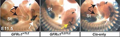
Enomoto first devised an elegant experimental system to make it possible
to generate mice specifically lacking RET-independent GFRα1. The study
of GFRα deficiencies in vivo is dogged by the lethality of the phenotype,
in which the absence of enteric neurons and functioning kidneys results
in death soon after birth. In vitro studies and the proximity of RET-independent
GFRα and RET-expressing cells in some developmental regions, however,
have prompted strong speculation that GFRα might be able to operate even
in the absence of RET indigenous to the cell. It has been suggested that
this might take the form of either trans signaling, in which
the GFRα receptor captures diffusible GFLs and presents them to a neighboring
RET-expressing cell, or through a separate signaling mechanism mediated
by GFL-activated neural cell adhesion molecules (NCAMs). Given this body of work showing the likelihood of a physiological role
for RET-independent GFRα1 activity, Enomoto et al. decided to test whether
the in vitro evidence would be borne out in living mice. The team showed
that mice homozygous for a transgene deleting an important segment of
the GFRα1 gene died, while heterozygotes (which carried only a single
copy of the transgene) were healthy and fertile. On comparing specific
embryonic regions in hetero- and homozygous mice, they found associations
between RET-expressing and RET-independent GFRα1 cells in kidney, enteric
and motor neurons, as well as the expected disturbances in development.
However, when they next generated mice that were only capable of expressing
GFRα1 only in the RET-expressing cells (by cloning GFRα1 cDNA into
a region under the control of the Ret promoter and crossbreeding
the resulting animals with GFRα1 heterozygotes), they were surprised
to discover the mice were born healthy and free of any evident developmental
defects in the kidney or nervous system. They found no trace of GFRα1 mRNA in non- Ret -expressing cells in these mice (which they
named Cis-only mice, for their lack of trans signaling), while
GFRα1 transcripts were detected as expected in RET-positive cells, proving
that the conditional expression scheme had worked. Analysis of individual regions known to be susceptible developmental
failure on loss of GFRα1 function, such as the kidneys, motor and enteric
neurons and certain parts of the central nervous system during development
and following injury, showed that Cis-only mice develop and regenerate
structures that are both morphologically normal and fully functional. Investigating the second question of a possible alternate RET-independent
GDNF receptor complex thought to involve neural cell adhesion molecules,
they next examined Cis-only mouse olfactory bulbs. These bulbs are reduced
in size in NCAM-deficient mice as the result of impaired migration of
neural precursors through a zone called the rostral migratory stream and
swell with cells that have failed to reach their normal destination; this
phenotype is seen only weakly in mice lacking GFRα1 (which is thought
by some to regulate NCAM-mediated cell adhesion), but not in mice lacking
RET. Again, the Cis-only mice showed no discernible differences from wild
type.
|
||
|
||
[ Contact ] Douglas Sipp : sipp@cdb.riken.jp TEL : +81-78-306-3043 RIKEN CDB, Office for Science Communications and International Affairs |
| Copyright (C) CENTER FOR DEVELOPMENTAL BIOLOGY All rights reserved. |