| RIKEN Center for
Developmental Biology (CDB) 2-2-3 Minatojima minamimachi, Chuo-ku, Kobe 650-0047, Japan |
| Shisa serves double duty in the control of head development | ||
January 28, 2005 – The network of regulatory factors involved in the development of the vertebrate head is an exquisitely tuned system of almost baroque complexity. During early embryogenesis, arrays of activating and inhibitory factors work to mold an initially unstructured clump of cells into an increasingly recognizable body with a distinct back, belly, head and tail. The development of the head is enabled and guided by the activity of a region of cells called the organizer, which secretes inhibitors of multiple regulatory molecular pathways that affect the axial orientation and growth of the embryo; in a sense, the organizer acts as a defense system against a host of suppressors that would otherwise frustrate the development of the nascent head. The activities of these secreted factors cross the boundaries of single cells, but it has been suggested that additional regulators of head development may be present and act autonomously within individual cells as well.
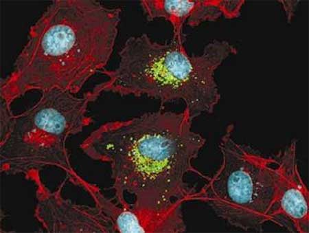
The Wnt and FGF signaling pathways are families of multifunctional molecules that work in cell growth and differentiation and the patterning of tissues and anterior-posterior body axes. As with proteins in secretory pathways, Wnt and FGF ligands and their cognate receptors are processed by the endoplasmic reticulum (ER) following translation, where they are folded into their functional configurations and undergo modifications such as the addition of sugars to their peptide chains. Only after attending this “finishing school” are the proteins ready for transport to the surface to serve as transmembrane receptors or be released into the extracellular space. Now, in an article published in the January 28 issue of Cell, Akihito Yamamoto of the Laboratory for Vertebrate Body Plan (Shinichi Aizawa, Group Director) and colleagues at the RIKEN Center for Developmental Biology (Kobe, Japan) report the identification of a novel factor, Shisa, which blocks Wnt and Fgf signaling at the endoplasmic reticulum by a mechanism previously unencountered in a developmental setting. shisa mRNA is expressed in both the head organizer and the anterior neuroectoderm, the region from which the head emerges, in embryos of the African clawed frog (Xenopus laevis), and has homologs in other vertebrates including mouse, zebrafish and human. Shisa encodes a secretory pathway protein with no known protein motif. In an early exploration of its role, Yamamoto found that its overexpression causes an expansion of the anterior territory, a property that earned the molecule its name. (The shisa is a form of sculpture common to Okinawa, representing a guardian lion-dog with a large head.) Further investigations of shisa function, using an experimental system known as animal cap assay, indicated that while Shisa has no discernible neuralizing activity, it acts as an anteriorizing agent that antagonizes both Wnt and FGF signaling. Loss of Shisa function revealed that Shisa is an essential factor for the proper formation of the head, especially in the presumptive head ectoderm during gastrulation. These initial in vivo studies suggested that Shisa is a secreted protein with a role as an inhibitor of Wnt and FGF signaling, which would have made it yet another factor discharged by the cells of the head organizer. But a closer examination of Shisa’s distribution within the cell revealed a distinct pattern of localization to the endoplasmic reticulum. Furthermore, in epistatic analysis, Shisa cell-autonomously inhibited signaling in receptor expressing cells. When the team watched the activity of Frizzled (Fz, a Wnt receptor) in cells also expressing Shisa, they found that both proteins remained within the endoplasmic reticulum (Fz is normally transported to the cell’s surface membrane). Looking for possible interactions, they found that Shisa specifically engages with a lower molecular weight species of Fz8, which they reasoned might be an immature version of the functional receptor protein, prior to its processing by the ER. Cells in which Shisa function was lost demonstrated promoted surface expression of Fz and increased responsiveness to stimulation by Wnt ligands, consistent with the idea that receptor activity had been enhanced. These findings led Yamamoto et al to conclude that Shisa works by holding the unfinished Wnt receptor at the endoplasmic reticulum, interfering with its maturation and resulting in its degradation before it can establish itself as a functioning unit within the cell membrane. Parallel investigations revealed similar ER co-localization with and inhibitory effects on an FGF receptor (FGFR), adding new novelty and interest to the story, as FGF signaling has posteriorizing activity, but is also required for neural development. It is not yet known how Shisa balances the repression of posteriorization while allowing FGF-driven neural induction in the anterior region to take place, a question that invites further study. The protein structure of Shisa and the exact mechanism by which it binds to and exerts its inhibitory effects on two structurally dissimilar receptor proteins also remain unknown. Indeed, the identification of a molecule capable of regulating a pair signaling pathways as central as Wnt and FGF by a novel mechanism seems destined to present new challenges of explication to cell and developmental biologists; as in any worthy exploration, the ultimate goal is not arrival, but a pushing back of the horizon.
|
||
|
||
[ Contact ] Douglas Sipp : sipp@cdb.riken.jp TEL : +81-78-306-3043 RIKEN CDB, Office for Science Communications and International Affairs |
| Copyright (C) CENTER FOR DEVELOPMENTAL BIOLOGY All rights reserved. |