| RIKEN Center for Developmental Biology (CDB) 2-2-3 Minatojima minamimachi, Chuo-ku, Kobe 650-0047, Japan |
| Let there be light-sensing cells | ||||
August 9, 2005 – Humans are visual animals. Our reliance on sight is immediately apparent on considering the impact the loss of that ability can have on a person’s range of everyday functions. The eye’s marvelous ability to sense light depends on the function of a population of cells in the retina, known as photoreceptors, which comprises the familiar “rod” and “cone” shaped cells responsible for contrast and color vision. And, as is generally true of neural cells, photoreceptors regenerate poorly or not at all, making retinal degenerative disorders, in which this specific group of cells is affected, both intractable to currently available therapies and promising candidates for treatment by regenerative medicine. In this emerging field of medicine, clinicians and researchers seek to replace cells that have been damaged or lost and so restore physiological function, as well as to develop systems for studying human diseases by modeling them in vitro. But access to pure, safe and plentiful supplies of specific types of cell remains one of the primary hurdles to be overcome before the promise of this young discipline can be realized. Now, in the August 2 online edition of the Proceedings of the National Academy of Sciences, Yoshiki Sasai (Group Director, Laboratory for Organogenesis and Neurogenesis) and colleagues at the RIKEN Center for Developmental Biology and Kyoto University report the world-first achievement of a method for inducing the differentiation of neural retinal precursors and photoreceptors from embryonic stem cells (ES cells) in mouse.
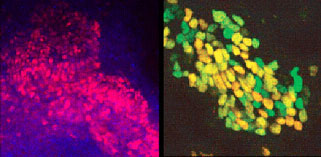
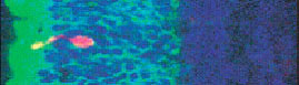
The Sasai group has published several previous studies showing the successful differentiation of a range of specialized neurons, including dopaminergic, enteric and forebrain, from ES cells. Although these pluripotent stem cells are, by definition, capable of giving rise to any type of cell in the body, limiting that potential to the generation of only a single type of cells requires close and detailed guidance. In the laboratory, this takes the form of highly controlled methods of ES cell culture. In this latest work, Sasai et al tweaked a protocol they had previously developed, called SFEB (for Serum-free Floating culture of Embryoid Body-like aggregates), which they showed earlier this year could yield neural differentiation at efficiencies of up to 90%. Knowing that photoreceptors arise from a population of neural retinal precursor cells identifiable by specific patterns of gene expression, the group first looked for ways of mimicking in vitro the differentiation pathway followed by the developing embryo. After much experimentation with various combinations of culture conditions and growth factors, Hanako Ikeda (now at the Kyoto University Hospital Translational Research Center) discovered that by treating floating aggregates of ES cells with activin, fetal calf serum and a pair of factors (Dkk1 and LeftyA) known to drive undifferentiated cells to a neural fate, she was able to induce their differentiation into cells bearing all the molecular hallmarks of neural retinal precursors at efficiencies as high as 16%. They dubbed the new method SFEB/DLFA. Further tests yielded even more exciting results. By culturing together with retinal cells from embryonic mice, the neural retinal precursors gave rise to large numbers (around 14% of entire cultured aggregate) of cells expressing rhodopsin and recoverin, two molecules known to be specific to photoreceptors in vivo. On culture with explants of embryonic neural retina, these ES cell-derived rhodopsin-positive cells behaved as photoreceptors would be expected to, integrating into sites in the neural retina explant where these cells normally appear.
This set of findings represents a significant advance, both as a demonstration of the importance of microenvironmental influences on embryonic cell decision-making, and as a proof-of-concept of a much sought after means to selectively induce photoreceptors from ES cells in culture. For patients and physicians confronting a range of disorders that damage the retina and threaten eyesight, such as age-related macular degeneration and the complications of diabetes, this should be welcome news indeed. Much research remains to be done before ES cell-derived photoreceptors can be brought to bear on human health problems but, as a first step, this work by Sasai and colleagues exemplifies the noblest aspirations of any science: to shed light and to offer hope. |
||||
|
||||
[ Contact ] Douglas Sipp : sipp@cdb.riken.jp TEL : +81-78-306-3043 RIKEN CDB, Office for Science Communications and International Affairs |
| Copyright (C) CENTER FOR DEVELOPMENTAL BIOLOGY All rights reserved. |