| RIKEN Center for Developmental Biology
(CDB) 2-2-3 Minatojima minamimachi, Chuo-ku, Kobe 650-0047, Japan |
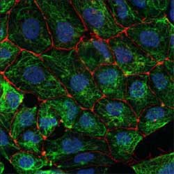
Now, in a series of experiments using mouse ES cells, the Laboratory for Stem Cell Biology (Shin-Ichi Nishikawa; Group Director) has demonstrated a means for inducing their differentiation both to visceral endoderm and, via an intermediary mesendodermal step, to definitive endoderm and mesoderm. This work now makes it possible for the first time to track these differentiation events in culture and to steer differentiation toward each of these lineages at high efficiency and to develop purified populations of cells sorted by their expression of surface antibodies. The studies have been described in a pair of reports published separately in the October issue of Development and the December issue of Nature Biotechnology. Looking at patterns of gene expression in the region of the early embryo from which the endoderm derives, they identified goosecoid (gsc) as a factor likely to be involved in endodermal differentiation and surmised it might have utility as an endodermal marker. The group next knocked-in a version of the gene tagged with a green fluorescent protein and experimented with the cells in vitro until they had a method of inducing differentiation that yielded a population of cells nearly all of which glowed green (signalling that their expression of gsc.) Next they screened the gsc+ colonies and determined that cells positive for goosecoid, e-cadherin and PDGFRa (gsc+ECD+aR+) possessed the twofold potency to differentiate into both mesoderm and endoderm characteristic of mesendoderm in other species. Even more intriguingly, this mesendodermal population could be subdivided into gsc+ECD-aR+ and gsc+ECD+aR- complements, which the group found differentiated into mesoderm and definitive endoderm, respectively. Turning to cells in which the goosecoid gene was not fused to express fluorescent protein, they demonstrated that the ECD and aR did their job equally well in genetically unmodified cells. Armed with this knowledge, the lab subsequently created a cell line in which the goosecoid locus bore the gene for gfp and huCD25 was introduced into the sox17 locus (sox17 is a known marker for extraembryonic visceral and definitive endoderm). This double knock-in enabled them to sort the definitive (gsc+sox17+) from the visceral (gsc-sox17+) fractions as the cells differentiated and, from there, to develop culture media best suited to nudging cells toward either of these lineages.Comparing gene expression in definitive and visceral endoderm, they identified seven surface proteins whose patterns of expression differed between populations. One of these molecules, CXCR4, is expressed specifically in definitive endoderm, meaning that by creating a monoclonal antibody against this protein, Nishikawa et al were able to track differentiation in unadulterated ES cells as well and to obtain pure populations of definitive endoderm by cell sorting.
|
||||
|
||||
[ Contact ] Douglas Sipp : sipp@cdb.riken.jp TEL : +81-78-306-3043 RIKEN CDB, Office for Science Communications and International Affairs |
| Copyright (C) CENTER FOR DEVELOPMENTAL BIOLOGY All rights reserved. |