| Tlx blazes a trail for blood vessel growth |
 |
 |
February 1, 2006 – Oxygen is a critical fuel for cellular activity, and cells respond actively to changes in their oxygen supply to ensure that demand is met. Hypoxia, the shortage of oxygen, triggers mechanisms in many tissues that kick-start the process of angiogenesis, in which new blood vessels sprout from the existingvasculature, laying down new pathways for oxygen-carrying blood to reach underprovided regions. This process is important in normal development as well, as the body needs to ensure that the metabolic needs of its tissues and organs are met.
The mouse provides a useful model for studying this kind of developmental angiogenesis in a mammal, as the vascular development of the retina forms postnatally, following an influx of astrocytes induced by the retinal neurons. These astrocytes secrete factors that initiate vasculogenesis, such as VEGF, as well as fibronectin, an extracellular protein that forms a trellis-like meshwork along which the nascent vascular sprout crawls. While the hypoxia-inducible regulation of VEGF has been well characterized in general, the mechanisms by which astrocytes respond to oxygen debt and satiation have largely been a mystery.
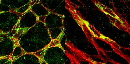
In contrast to the filamentous extracellular deposition of fibronectin proteins (green) around the astrocyte networks (red) in normal retinas (left), fibronectin proteins were absent in the extracellular spaces, but retained in the cytoplasm of retinal astrocytes in Tlx knockout mice (right). |
Now, in a study published in the February issue of The Journal of Clinical Investigation, Akiyoshi Uemura and colleagues in the laboratory for Stem Cell Biology (Shin-Ichi Nishikawa; Group Director), Regeneron Pharmaceuticals and the Salk Institute (USA), report that astrocyte behavior is mediated by a molecular switch that alters fibronectin assembly in response to changes in the cell’s oxygen supply. This regulator, Tlx, is a transcription factor expressed specifically in proangiogenic retinal astrocytes and which down-regulates dramatically on contact with blood vessels. Its specificity to unvascularized regions was linked to converse activity by another factor, GFAP (which is expressed only in established blood vessels), while mapping neatly to the expression patterns of VEGF and fibronectin in proangiogenic astrocytes. Interestingly, while both Tlx and VEGF appeared to respond to changes in oxygen concentration, fibronectin mRNA continued to be expressed even when there was a physiological surplus of oxygen.
Curious about the function of the Tlx gene product, Uemura found that, in Tlx-/- mutants, while astrocyte proliferation was only moderately affected, the astrocyte network failed to form in the retina and, as a result, blood vessels were entirely absent. The group conjectured that this may be due to defects in either cell motility or adhesion which prevent the advance of the growing vasculature, a hypothesis that was supported by their observation that ectopic vessels derived from the hyaloid vasculature (a fetal vascular system in the eye) that were attracted to astrocyte-rich areas of the retina failed to align themselves along the extant astrocyte network once on entering the mutant retina. Their speculation was proven right when they checked fibronectin expression and found that the null mutants’ astrocytes produced only scarce amounts of this scaffold-building protein, unlike the profuse networks laid down in wild type.
On the implications of his work, Uemura comments, “There are a number of human visual impairments, including diabetic retinopathies and infant R.O.P. (retinopathy of prematurity) which are related to defects in the vasculature. It’s exciting to think that this work on a developmental system might contribute to what we know about vessel growth in the human retina, and that this knowledge of the role of the astrocytic scaffold might one day lead to a therapy.”
|
