RIKEN Center for Developmental Biology (CDB)
2-2-3 Minatojima minamimachi, Chuo-ku, Kobe 650-0047, Japan


Cv2 boosts BMP signal
PDF Download


November 28, 2006 – Potent signaling factors, such as the bone morphogenetic protein (BMP) family of molecules, need to be regulated by a system of checks and balances to ensure that they operate at precisely the right time, location and intensity during development. Their ability to effect fundamental changes in cellular differentiation means that this regulation is frequently negative, preventing them from working in inappropriate contexts; the BMPs, which have a powerful ventralizing effect, are famously counteracted by a set of BMP antagonists, such as chordin, in the dorsal side of the embryo. But positive regulation plays an important role as well, when an amplified signaling is called for at a particular site or stage. In the fly, the chordin homolog Sog is believed to upregulate BMP in some contexts, but in vertebrates, the case for BMP potentiation is less clear.

Now, in an article published in the journal Development, Makoto Ikeya and colleagues in the Laboratory for Organogenesis and Neurogenesis (Yoshiki Sasai; Group Director) report a gene, Cv2, which acts as a local enhancer of BMP signaling in mouse development. Named for its homology to the Drosophila gene crossveinless 2, which enhances BMP activity in the formation of cross-veins in the fly wing, mouse Cv2 works to amplify BMP signaling in the development of a number of tissue and organ systems, including bone, cartilage, kidney and eye.

The deletion of one copy of BMP4 strongly and cooperatively enhanced the vertebral defect of mCv2-/- mutants, causing reduction of the vertebral body in size and of its ossification and suppression of vertebral arch development. Moreover, the reduction of the BMP4 gene dosage synergistically increased the frequency of the microphthalamic phenotype.


The study began with a database search that identified several mouse genes, including Cv2, related to the BMP regulators chordin and kielin. Ikeya et al generated knockout mice carrying a null mutation for Cv2 to study its role in murine embryogenesis. Although the appearance of these mutants was not dramatically different than that of wild type animals, they showed a range of embryonic phenotypes and all died in the perinatal period. Multiple defects were observed in skeletal and cartilage development, resulting in a shortened trunk. Affected regions included the bones of the spine, ribs, skull and limbs, and cartilage in the pharynx and trachea.

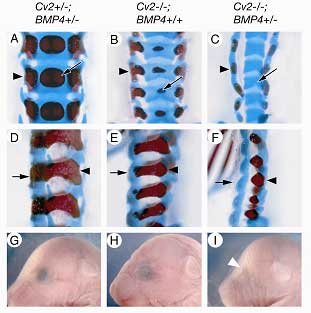
The pattern of these defects suggested that the loss of Cv2 prevents the differentiation of precursor cells derived from the sclerotome, a subset of cells that migrates out from the somites to give rise to the vertebrae. But analysis of gene expression by RT-PCR in tissues from the embryonic trunk indicated that the effect was not due to a simple, generalized reduction in BMP gene expression. Tests performed in cultured cells pointed instead to a role for Cv2 upstream of or at the level of the BMP molecular receptor.

Interested in the nature of the interaction between Cv2 and BMP signaling, Ikeya next generated a series of mutants engineered to full or partial deletions of both genes, and found that in mice carrying a null mutation for Cv2, the loss of a single copy of the gene BMP4 significantly intensified the defects in both vertebral and eye development, suggesting a cooperative relationship between the genes.

BMPs are known to be required for renal development as well, so the group next looked at the kidneys in the Cv2 mutants, and found that they were smaller and contained fewer renal glomeruli than normal. Taken together with the fact that the kidney mesenchyme normally strongly expresses Cv2, these findings pointed to an essential role for the gene in kidney development. On testing the effect of the loss of function of both Cv2 and the structurally similar kielin-chordin related protein (Kcp) on nephrogenesis, Ikeya et al found that the phenotype was more severe in the double mutant, indicating that the genes work together in the regulation of kidney development. Interestingly, the skeletal defects were unaltered in the Cv2/Kcp mutants.

This thoroughgoing tissue-by-tissue analysis of the effects of loss of Cv2 function reveals that the gene is required in highly specified processes to enhance BMP signaling – while vertebrae, eye and kidney are all affected, other important BMP-dependent regions, such as some skeletal components and the dorsoventral patterning of the neural tube, are not. Whether this context-sensitive pro-BMP of Cv2 is achieved by enhancing the association of BMP ligands and receptors, competition with BMP antagonists, controlling the sequestration of BMP proteins in the matrix, protecting them from protease degradation, or some other mechanism, remains unknown.


Link to article http://dev.biologists.org/cgi/content/abstract/dev.02647v1


Copyright (C) CENTER FOR DEVELOPMENTAL BIOLOGY All rights reserved.