| RIKEN Center for Developmental Biology (CDB) 2-2-3 Minatojima minamimachi, Chuo-ku, Kobe 650-0047, Japan |
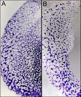
Now, in an article published in the journal Blood, Fumie Nakazawa and Hiroki Nagai in the Laboratory for Early Embryogenesis (Guojun Sheng, Team Leader) show an important function for the FGF receptor FGFR2 in regulating cellular fate during primitive hematopoiesis. Investigating patterns of gene expression and FGF gain and loss of function in the embryonic chicken, Nakazawa and Nagai demonstrate that FGF signals potently inhibit hemoglobin expression (a hallmark of hematopoiesis) and upregulate endothelial markers, while loss of FGF signaling results in ectopic blood formation.
Nakazawa and Nagai next tried the converse experiment, using beads soaked in a molecular inhibitor of FGF signaling, and found that primitive blood cells began to form ectopically in both grafts and intact embryos, while the endothelial cells that give rise to the vasculature failed to form. The inhibitory agent used was a general inhibitor of all FGF receptors (FGFRs), of which there are four in the chick, so they next examined the patterns of gene expression for each of the FGFRs in situ and found FGFR2, which is expressed in endothelial and absent in blood cells, to be the likeliest candidate for a specific regulator of primitive hematopoiesis in the extraembryonic mesoderm. Zeroing in on this target, they used electroporation to introduce a constitutively active version of FGFR2 into early embryos and compared their subsequent cellular composition with that in control embryos. In the control sample, roughly one-third of the cells expressed ρ (indicating a blood fate), another third were of endothelial lineage, with the remainder being of other cell types. In the FGFR2 embryo, however, less than 3% of the cells were found to express ρ, and more than 80% differentiated into endothelium. When morpholinos were used to block the function of the endogenous FGFR2, the opposite effect was seen. Having demonstrated a strongly inhibitory effect for FGFR2 on primitive hematopoiesis, the Sheng lab sought to put the receptor in context within the many molecules signals known to impact on blood differentiation. Inhibition of VEGF signaling, another pathway implicated in blood and endothelial development, yielded no appreciable effect on the expression of either ρ or the endothelial marker, Lmo2. But beads soaked in the FGF ligand FGF4 had a negative effect on the expression of Gata1, a member of a second set of genes known to be expressed in the blood lineage immediately prior to the onset of globin expression. And in FGF-inhibited embryos, Gata1 expression was found to be upregulated and expanded in patterns similar to those seen for ρ. This work by Nakazawa, Nagai and colleagues points to a role for the receptor FGFR2 in inhibiting primitive blood differentiation, but it remains to be seen which FGF ligand is at work in this process. And while FGFR2 has now clearly been shown to play an important part, the intricate web of molecular pathways that establish and maintain the tight balance between blood and vascular differentiation in clusters of cells during early development continues to hold a few secrets. “We know very little of how blood island cells are set aside initially and how the differentiation of smooth muscle cells (which are not derived from blood islands) is coupled with that of the endothelial cells,” says Sheng. “And it is of tremendous importance, both for us and for people working in stem cell biology, to know how a very small percentage of blood island cells remains undifferentiated. |
|||||
|
|||||
 |
| Copyright (C) CENTER FOR DEVELOPMENTAL BIOLOGY All rights reserved. |