| RIKEN Center for Developmental Biology (CDB) 2-2-3 Minatojima minamimachi, Chuo-ku, Kobe 650-0047, Japan |
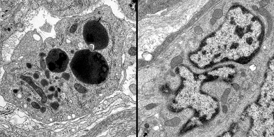
“This discovery that an unconventional form of neuronal cell death is triggered by inactivation of GFRα1 in the distal colon is exciting for us,” says Uesaka, “as it may provide us with valuable insights into the pathogenesis of Hirschsprung's disease, a disease in which ganglia fail to form in the distal colon in humans.” Early in ENS development, the GFRα1 receptor is expressed in the majority of enteric neuronal progenitors differentiating neurons throughout the gut, but its distribution gradually shifts over time until it is predominantly expressed in enteric neurons in the colon. This caught the team’s attention, because GDNF is also detected at particularly high levels in the colon and caecum during later stages of embryogenesis. Uesaka was interested in studying the role of GFRα1, so to avoid the total loss of enteric neurons characteristic of the unconditional knockout of the gene, he painstakingly generated a line of mice in which the function of GFRa1 could be excised contingently and cells carrying an inactivated allele traced by the expression of green fluorescent protein. When he looked at embryos in which GFRa1 was inactivated at day 15.5 (by which point neuronal GFRa1 expression is nearly confined to the colon), he found that the normal pattern of lower gut innervation was completely disrupted, with a massive loss of enteric ganglia. Tests of the conditional knockout at other developmental time points showed that this effect was time-dependent; embryos in which GFRa1 was excised earlier in development showed defects in enteric neuronal proliferation, but not survival. Curiously, however, the conditional E15.5 knockouts showed none of the telltale signs of apoptosis; tests revealed no significant upregulation of typical cell death markers such as activated caspase, nor the DNA fragmentation apoptotic cells undergo (as revealed by TUNEL assay). Following up on this startling observation, the Enomoto lab turned next to the proaptototic factor, Bax, but found that Bax deficiency had little influence on enteric neuron death induced by GDNF deprivation in culture. And, while a number of abnormal morphological features were seen in the nuclei cells in colonic neurons of conditional GFRa1 knockout embryos, they showed none of the nuclear hallmarks of apoptosis, such as globular or crescent-shaped chromatincondensation, nor did dying cells appear to be succumbing to autophagy, as many apoptotic cells do. In addition to demonstrating that GDNF is a survival factor for enteric neurons, a role it plays in many other arenas of neurodevelopment, Uesaka’s study points to an intriguing new form of physiological cell death, which may be important in other developmental contexts and neuronal pathologies as well. Enomoto notes of the study’s medical implications, “We think that the possible links to the etiology of Hirschsprung’s disease are especially interesting, as we know that RET, the signaling component of the GDNF receptor complex, is most frequently mutated in those patients. It will certainly be worth looking into whether and how this novel cell death mechanism might apply in humans.” |
|||||
|
|||||
 |
| Copyright (C) CENTER FOR DEVELOPMENTAL BIOLOGY All rights reserved. |