| RIKEN Center for Developmental Biology (CDB) 2-2-3 Minatojima minamimachi, Chuo-ku, Kobe 650-0047, Japan |
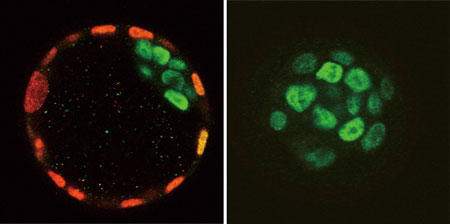
Noriyuki Nishioka, Shinji Yamamoto and others in the Laboratory for Embryonic Induction (Hiroshi Sasaki; Team Leader) have now shown that the transcription factor Tead4 is required for trophectoderm development, in a manner that suggests it functions upstream of Cdx2. Their findings, which were published in the journal Mechanisms of Development, shed new light on the genetic regulation of the earliest cell specialization event in mammalian embryogenesis. The Sasaki lab’s study began with the generation of mice with a homozygous deletion of the Tead4 gene, following on their previous work that showed roles for Tead-family genes in the regulation of Foxa2, a transcription factor known to regulate the development of the midline signaling centers controlling post-implantation development. They were surprised to find that the knockout embryos died at extremely early stages in development, prior to the implantation of the blastocyst into the uterine wall. This unexpected early lethality prompted Nishioka and Yamamoto to re-examine the expression of Tead4, and the related genes, Tead1, -2, and -3, in pre-implantation mouse embryos. Using RT-PCR to detect Tead transcripts, they found that Tead4 expression switched on by the 4-cell stage; two other Tead genes, Tead1 and Tead2, were also expressed. They next used immunohistochemistry to attempt to detect the various Tead proteins, and found both Tead1 and Tead4 in all blastomeres (as the individual cells of the early embryo are called), as well as in the cells of trophectoderm and the inner cell mass slightly later in development. They turned next to the trophectoderm, as this tissue is also a critical requirement for normal blastocyst development. Testing for Cdx2 expression, which is upregulated in the trophectoderm in wildtype embryos and required for TE lineage specification, they found that Cdx2 is only faintly expressed in the Tead4 mutant embryo after the first several rounds of cell division, and not at all in subsequent stages. This was true of other TE-specific genes, such as Eomes and Fgfr2, as well. Cdx2 functions in the pre-implantation embryo by repressing the expression of Oct4. When Nishioka and Yamamoto checked Oct4 expression, they found that it was expressed throughout the late blastocyst, indicating that the entire embryo had adopted an inner cell mass fate. Given that the ICM can serve as a source for embryonic stem (ES) cells, the team tried to establish an ES cell line using Tead4 mutants, and succeeded in establishing three ES-like cell lines, one of which was capable of forming colonies in culture and differentiating into all three germ layer lineages in vitro, suggesting that Tead4 is not required for development of embryo proper. “The finding that trophectoderm completely fails to form in Tead4 mutants provides an important clue for us to better understand early development in mammals,” says Sasaki. “But the important question of how this gene, which is expressed in every cell in the embryo, can prompt trophectodermal differentiation in only a subset of those cells still needs to be addressed.” |
|||||
|
|||||
 |
| Copyright (C) CENTER FOR DEVELOPMENTAL BIOLOGY All rights reserved. |