| RIKEN Center for Developmental Biology (CDB) 2-2-3 Minatojima minamimachi, Chuo-ku, Kobe 650-0047, Japan |
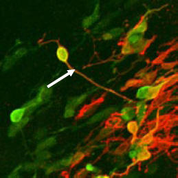
A report by Fumitaka Osakada, Hanako Ikeda and colleagues in the Laboratory for Retinal Regeneration (Masayo Takahashi; Team Leader) now makes an important stride toward realizing that dream. Their study, conducted in collaboration with clinicians and scientists from Kyoto University, and the CDB Laboratory for Organogenesis and Neurogenesis (Yoshiki Sasai; Group Director) and published in Nature Biotechnology, reports the high-efficiency generation of rod and cone photoreceptors and pigmented epithelium from mouse, primate and human embryonic stem cells under fully characterized culture conditions. The Takahashi lab and others had previously developed methods for inducing the differentiation of retinal neural progenitors, but these relied on co-culture with embryonic retinal tissue or other confounding factors, and generally produced retinal neural cells at unacceptably low efficiencies. Working first with mouse embryonic stem (ES) cells, Ikeda sought to identify factors capable of spurring photoreceptor differentiation in a more reliable and efficient manner. After isolating progenitor cells and confirming their competence for further differentiation to retinal precursors (as shown by their expression of the marker gene Crx), the Takahashi team found that the γ-secretase inhibitor DAPT steered the mitotic progenitors to adopt a postmitotic precursor fate. They next tested a panel of factors implicated in photoreceptor differentiation on the Crx+ precursor cells, and found that a combination of aFGF, bFGF, taurine, Shh, and retinoic acid in retinal culture medium prompted nearly 30% of the precursors to begin expressing the light-sensitive opsins characteristic of either rod or cone photoreceptors. One drawback of the method used for inducing differentiation from mouse ES cells is that it involves the use of fetal bovine serum, which problematizes its use for clinical applications. Seeking a workaround, Osakada turned next to a chemically defined medium for use with ES cells from cynomolgus monkeys and found that the serum free floating culture of embryoid body-like aggregates (SFEB) system worked for primate cells as well, without requiring the use of bovine serum. They next tested a pair of factors that had been shown to promote retinal-lineage differentiation – the Wnt-antagonist Dkk1 and the nodal-antagonist Lefty-AS – and found that in combination, these were highly effective in boosting the differentiation of retinal pigmented epithelium, as marked by the factor Mitf. Osakada et al. further found that by treating retinal progenitors derived from monkey ES cells with retinoic acid and taurine, two of the factors used in the mouse ES study, they could strongly promote differentiation of Crx-positive postmitotic precursors, and subsequently of rod and cone-like cells expressing opsins appropriate to their putative cell types. In a final set of experiments, the Takahashi lab assessed whether their findings using mouse and primate ES cells could be replicated using human embryonic stem cells as well. Using the SFEB culture (originally developed by the Sasai group) optimized for use with human ES cells, they found that as in the monkey ES cell tests, the combination of Dkk1 and Lefty-A caused a significant number of the undifferentiated cells to assume a retinal pigmented epithelial fate, as determined by morphology and gene expression. Differentiation of Crx+ precursors into photoreceptor-like cells using taurine and retinoic acid also proved successful, although at slightly lower efficiency than in monkey cells, establishing this new technique as a potentially important tool for generating human photoreceptors for use in drug screening and future regenerative medical applications. “Now we have the cells that might one day be useful for retinal cell transplantation therapy, but it needs to be remembered that these are only some of the raw materials. We will still need to establish methods for purifying them, and getting them to function in vivo after transplantation,” cautions Takahashi. “We also need to recognize the differences in closeness to clinical application between retinal pigmented epithelial cells and photoreceptor cells. We now need to think practically about how to translate this basic research, and the hurdles to that lie ahead.” |
|||||
|
|||||
 |
| Copyright (C) CENTER FOR DEVELOPMENTAL BIOLOGY All rights reserved. |