| RIKEN Center for Developmental Biology (CDB) 2-2-3 Minatojima minamimachi, Chuo-ku, Kobe 650-0047, Japan |
May 12, 2009 In vertebrates, the eye develops from a single patch at the headward end of the neural plate, which later splits into two, allowing a pair of eyes to form from optical vesicles that bulge outward from the sides of the developing brain. The initial bisection of the eye field is achieving by signaling from the neighboring prechordal mesoderm, but how the subsequent shaping and fine-tuning of the resulting halved regions is directed is not well understood.
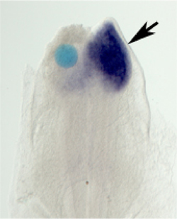
Now, in a report published in Developmental Biology, Michael Teraoka and colleagues in the Laboratory for Sensory Development (Raj Ladher; Team Leader) describe how a region of paraxial mesoderm works latterly to refine and restrict the eye field after its bisection in chick. The team found that signals from the rostral cephalic paraxial mesoderm help to limit the expression of eye field transcription factors through the action of the BMP signaling pathway. Teraoka began with experiments in which he removed various tissue regions at developmental stages corresponding to eye formation. When he extirpated paraxial mesoderm near the head, he saw the optical vesicles subsequently became enlarged, due to an increase in the total numbers of cells in these structures. As no effect was seen on cell proliferation, he surmised that the increase in the numbers of optical vesicle cells might be due to a misassignment of cell fate instead. Continuing to use a classic embryological approach, he next swapped rostral paraxial mesoderm from quails into regions of the embryonic chick head that do not normally feature mesoderm. He found that this affected eye development in the converse manner optic the cups were reduced in size and the lens vesicles were small or absent. The transplantation also affected the expression of genes associated with eye development, such as Pax6 and crystallin. Significantly, there was no effect on neural patterning in general, suggesting that the rostral paraxial mesoderm has an eye-specific role. This role, in turn, is specific to the rostral paraxial mesoderm as well; transplantation of other regions of quail mesoderm to the same part of the embryonic chick head had no effect on the development of the eye. Seeking a possible molecular mechanism behind the rostral paraxial mesoderms eye-inhibiting function, Teraoka focused on bone morphogenetic protein (BMP) signaling, which he found to be expressed in the paraxial mesoderm and, importantly, repressed in the axial mesoderm, which has no optic-inhibitory activity. He next implanted tiny beads soaked in BMP factors to test for effects on optic gene expression, and found that BMP4 caused reductions in several eye-related genes without affecting genes more generally involved in neural patterning. Knocking the BMP level down in vivo by shRNA had the opposite, pro-optic effect of increasing the size of the optic vesicles, which was mirrored by the antagonism of factors downstream in the BMP pathway as well. The team speculates that BMPs role may be mediated by its interplay with both canonical and non-canonical Wnt signaling, another pathway known to be important in eye development. I think that the most important aspect of this work is the finding that organ-generating fields are constantly being told what to do by their neighbors, says Ladher. In this way the final shape and position is formed so that the organ can carry out its function. |
|||||
|
|||||
 |
| Copyright (C) CENTER FOR DEVELOPMENTAL BIOLOGY All rights reserved. |