| RIKEN Center for Developmental Biology (CDB) 2-2-3 Minatojima minamimachi, Chuo-ku, Kobe 650-0047, Japan |
May 9, 2011 The process of programmed cell death known as apoptosis plays a critical role in many developmental events, such as the sculpting of digits in vertebrate limbs, in which the death of the cells that would otherwise form inter-digital webbing enables individual fingers and toes to be formed. The importance of the removal cells in such contexts in well known, but in recent years, we have begun to learn more about additional roles for apoptosis in the formation of tissues and organs.
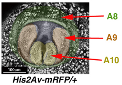
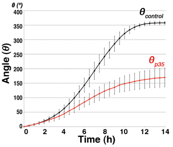
A new report by Erina Kuranaga, Team Leader of the Laboratory for Histogenetic Dynamics and colleagues reveals one such novel function for apoptosis in speeding the rotation of ring-like structures in the developing male reproductive system in the fruit fly Drosophila, which is necessary for its formation. This work, which was initiated at the University of Tokyo, is published in the journal Development. Kuranaga began by analyzing the part played by apoptosis in morphogenetic processes using the Drosophila system with its powerful genetics and decades-old store of research findings. In 1930, it was noted that in this fly the male reproductive structure known as the terminalia makes a full 360-degree rotation during development. More than half a century later, researchers observed that defects in apoptosis result in incomplete rotation, and consequently reduced reproductive function and shortening of the structural links with the terminalia. In her recent study, Kuranaga brought live imaging technology to bear on observation of the rotational kinetics of this system. The terminalia originate in the rostral section of the embryo, and comprises a ring-shaped region known as A8 that encircles the two inner regions A9 and A10. Kuranaga used fluorescent proteins to visualize the nuclei of cells in these domains, and observe their movements in living embryos. They found that rotation begins approximately 24 hours after the formation of the puparium, and takes around 12 hours to complete a full rotation. But when the team blocked the activity of caspases, which are responsible for inducing apoptosis, they found that while rotation began at the same developmental time point and continued over the same 12-hour period, it failed to complete a 360° turn. To gain a better understanding of how this happened, they quantitatively analyzed the terminalia rotation in the developing flies, and determined that in wildtype the process involves four distinct steps: initiation, acceleration, deceleration, and termination. In apoptosis-defective flies, however, the acceleration stage was not maintained, leading to incomplete rotation. They next conducted more sophisticated live imaging observations, allowing them to track the behaviors of individual cells. They saw that in the ring-like A8 domain the inner and outer populations of cells behaved differently the interior cells rotated 360° in synchrony with the cells of the A9 domain, but the outer cells only rotated half that distance, or 180°. Kuranaga also noted that the outer ring cells began their rotation at the same time that the inner cells were entering the acceleration phase, suggesting a link between these events. She also observed for the first time that apoptosis occurs in the A8 domain.
In embryos in which apoptosis was blocked, the inner cells in A8 moved as in wildtype, but the rotation of the outer cells failed to begin. By analyzing the frequency of apoptotic events and speed of rotation of outer cells in wildtype embryos as well, the team was able to show clear correlation between the two. Taken together, the findings suggest that apoptosis is required for the rotation of the outer ring, which in turn drives the acceleration of the inner ring of the A8 domain. And indeed, upregulation of the pro-apoptotic pathway resulted in faster rotation of the terminalia. The outer ring seems to function as a kind of moving walkway, says Kuranaga. When the inner ring steps onto it, its speed increases, allowing it to complete its rotation within the normal period. We are still working out how might apoptosis drive this, but suspect it may be related to the relief of some mechanical stress generated by cellular movements. | |||||||||
|
|||||||||
 |
|||||||||
| Copyright (C) CENTER FOR DEVELOPMENTAL BIOLOGY All rights reserved. |