| RIKEN Center for Developmental Biology (CDB) 2-2-3 Minatojima minamimachi, Chuo-ku, Kobe 650-0047, Japan |
December 12, 2011 The study of developmental processes frequently demands the ability to study how individual cells look and act. This has been bolstered in recent years by the development of technologies for fluorescence live imaging, which have made it possible to visually examine and monitor cells in living embryos of various species. Applications of live imaging techniques have been introduced in the mouse as well, but these are complicated the inability to simultaneously label multiple tissues using discrete markers, and the random integration of fluorescence genes, which makes it difficult to constrain sites and dosages of expression.
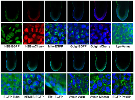
In a step forward for the field, the Laboratory for Animal Resources and Genetic Engineering (LARGE; Shinichi Aizawa, Laboratory Head) has developed 12 new mutant strains of mice engineered to conditionally express fluorescent markers at seven different cellular sites, such as nucleus or cell membrane. These new mice, engineered using conventional Cre-Lox recombination, express fluorescent proteins at levels sufficient for strong visualization, and enable dual labeling by different fluorescent signals at user-determined developmental sites and time-points. Published in Genesis, these novel mouse resources are now available for distribution from LARGE. In tackling the challenges posed by mouse biology for live imaging techniques, the lab focused on the ROSA26 locus, which enables introduced genes to be expressed nearly ubiquitously in mouse tissues, and can be mutated on both sites in a pair of chromosomes without causing abnormal phenotypes. They took advantage of these properties by introducing genes for fluorescent proteins into the ROSA26 locus and expressing them conditionally using the Cre-Lox system, which made it possible to engineer a series of mice specially adapted for use in live imaging studies. They kicked off the effort by fusing genes for any of six fluorescence signals, such as EGFP (green) and mCherry (red) with genetic markers of subcellular sites, such as nucleus and mitochondria, giving rise to a total of 28 reporter genes. Testing these for specificity and strength of expression narrowed the field to 16 candidates, which were then introduced by homologous recombination into the ROSA26 locus in mouse embryonic stem cells (ESCs) accompanied by a stop codon flanked by LoxP sequences, making it possible to conditionally switch on their expression only in cells expressing Cre. They next crossed these 16 reporter mouse lines with mice designed to express Cre constitutively, and examined their patterns of expression in E7.5 embryos. Twelve of the 16 expressed the reporter genes at appropriate sites and sufficient quantities to enable their use in imaging applications. For their next step, the team crossed mice that heterozygously expressed Venus (red) and mCherry (green) in the cell membrane and nucleus, respectively, thereby generating dual-labeled embryos, and making it possible for the first time to study the detailed movements of nuclei in epiblast stage embryos.
As a final test of the specificity of expression, they crossed mice expressing Venus (green) in the cell membrane with other designed to express Cre only in the notochord. Observing the resulting embryos at day 8.5 of development showed clear membrane expression of Venus in a tissue-specific manner.
The group published a second report in the same issue of Genesis, describing a proof-of-concept application of a dual-labeled line expressing mCherry in nucleus and EGPF in the plasma membrane. Again using the Cre-Lox system, they were able to achieve the specific expression of the genetic reporters for these two organelles only in fetal Sertoli cells, indicating the usefulness of these new resources in live imaging studies. | |||||||||
|
|||||||||
 |
| Copyright (C) CENTER FOR DEVELOPMENTAL BIOLOGY All rights reserved. |