| RIKEN Center for Developmental Biology (CDB) 2-2-3 Minatojima minamimachi, Chuo-ku, Kobe 650-0047, Japan |
June 14, 2012 Pluripotent stem cells such as embryonic stem cells (ESCs) and induced pluripotent stem cells (iPSCs) are valued for their ability to give rise to all the many cell types in the adult body. Since the first derivation of human ESCs in 1998, scientists have developed a variety of techniques for inducing these cells to differentiate into specific cell populations at reasonably high efficiencies. More recently, a number of reports have shown that mouse ESC can be prompted not only to form formless cell aggregates, but remarkably well-organized tissue-like structures in vitro, a feat that has been likened to the recapitulation of developmental phenomena in a culture dish. Now, Tokushige Nakano and colleagues in the Laboratory for Organogenesis and Neurogenesis (Yoshiki Sasai, Group Director) have shown that human ESCs exhibit that same capacity for generating self-organized, complex tissue structures with their report of an hESC-derived optic cup. Published in Cell Stem Cell in collaboration with Sumitomo Chemical, the report also describes the groups development of a cryopreservation technology capable of freezing these retinal tissues intact.
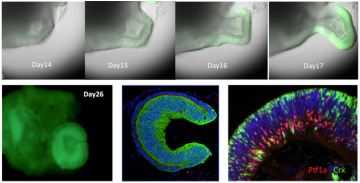
This represents the latest in a series of achievement in induced differentiation from ESCs by the Sasai group. Using a cell culture system (SFEBq) developed in the same lab, Sasai has previously shown controlled differentiation of a range of neuronal cell types including dopaminergic neurons, cerebellar Purkinje cells, as well as cortical, sensory and motor neurons. In recent years, the group has shown that mouse ESCs can be steered to self-organize into complex tissue-like structures involving multiple cell types, including cerebral cortex, optic cup, and pituitary hypophysis, revealing an intrinsic developmental capacity in these pluripotent cells independent of signals from the surrounding microenvironment. The most recent achievement built on the labs previous demonstration of optic cup formation from mouse ESCs, but required modifications to that protocol to account for difference in tissue size, number of cells, culture media, and the time points at which the growth factors that steer the process needed to be added. The optimized 22-26 day process began with a population of around 9000 hESCs, which formed retinal precursors after about two weeks in culture, after which the structure gradually remodeled to form the optic cup. Interestingly, the resulting structure was around twice the diameter of that derived from mouse ESCs, apparently reflecting the size differences in the embryos of the two species. When the neural retina was cut away from induced optic cups and cultured it showed even further differentiative capacity, giving rise to ganglion cells, photoreceptors and other cells types by day 40, and forming a laminar retinal structure resembling that of the adult retina by day 126. Intriguingly, Nakano also observed cone photoreceptors, a cell type not seen in mouse ESC-derived tissue. Recognizing that the long culture times required to give rise to these tissues could represent a pitfall for clinical use, the group also developed a system for pre-treating the optic cups prior to cryopreservation, allowing them to be frozen, stored, and thawed with minimal damage. The question of just how complex a structure we can obtain from embryonic stem cells is a fascinating one from the developmental perspective, says Sasai. We are hopeful that these findings may help to lay a foundation for a regenerative medicine in which intact organized tissues, not just groups of cells, can be developed for use in cell transplantation. |
|||||
|
|||||
|
|||||
 |
| Copyright (C) CENTER FOR DEVELOPMENTAL BIOLOGY All rights reserved. |