| RIKEN Center for Developmental Biology (CDB) 2-2-3 Minatojima minamimachi, Chuo-ku, Kobe 650-0047, Japan |
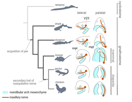
Dec 6, 2013 As its name implies, the trigeminal nerve has three major branches in vertebrates: a first division into the ophthalmic nerve and the maxillomandibular, which further bifurcates into the maxillary and mandibular nerves. The trigeminal governs sensation and muscle control in the face and jaw. In humans, it develops from a primordium just above the pharyngeal arches, from which the ophthalmic nerve diverges and extends toward the eye, and the maxillary and mandibular nerves toward their respective prominences. The structure of the jaw is conserved from fish to mammals, so the how its innervation patterns were acquired during evolution are a question of fundamental importance. In a comparative study of trigeminal development in diverse gnathostome taxa published in Journal of Morphology, Hiroki Higashiyama and Group Director Shigeru Kuratani of the Lab for Evolutionary Morphology have discovered that a branch of the trigeminal (called the nasopalatine nerve in human) innervates derivatives of the medial nasal prominence. Only diapsids, such as gecko and chicken, lacked a homolog of this nerve, suggesting it had been secondarily lost in this lineage.
Textbook accounts of maxillary nerve development typically state that it is localized to the maxillary prominence and goes on to innervate the entire upper jaw. But recent embryological studies have shown that the upper jaw arises not only from the maxillary prominence, but rather forms from the fusion of this primordial structure with the medial nasal domain, suggesting an apparent discrepancy between neural morphology and developmental pattern. The medial nasal prominence is the site of origin for the upper lip, anterior palate, and front teeth, and gives rise to the upper jaw (maxilla) by merging with the maxillary prominence. But despite the regions origins in a fusion with the medial nasal prominence, to which the maxillary nerve does not localize, the maxillary nerve appears to control the entire maxillary region. Interested in this conundrum, Higashiyama examined the distribution of trigeminal nerve branches in mouse. At day E10.5, he noted the clear projection of the maxillary nerve to the maxillary prominence, but by day E11.5 he saw that this nerve had begun to ramify rostrally. This branch, corresponding to the human nasopalatine nerve, follows the nasal septum to innervate the front teeth and soft palate, indicating that it is the nasopalatine that controls the medial nasal region in mammals. But what of other taxa? When Higashiyama looked at relatively closely related diapsids, such as reptiles and birds, he was surprised to find that the nasaopalatine was missing, with its function assumed by posterior facial nerves. This raised the question of which state was ancestral, and which derived. Higashiyama broadened his search to anamniotes, such as cartilaginous and bony fishes, and were happy to see that the maxillary and medial nasal prominences did not fuse, considerably simplifying the investigation. In these embryos, the maxillary nerve formed two branches, extending into both the maxillary and medial nasal territories, suggesting that the arrangement seen in mammals is common to all gnathostomes, and only secondarily absent in diapsids. When he looked at the lamprey, a jawless fish, Higashiyama found something even more interesting the nerve projecting to its round mouth corresponds to the nasopalatine, suggesting that it, not the maxillary, is the more ancient structure. Studies of vertebrate morphology often focus on hard structures, such as skeletal elements, but our bodies are mostly made up of softer bits, including the nerves we looked at in this study, says Kuratani. Similar to the trigeminal, the vagal nerve plays an important role in controlling enteric functions, but the relationship between this nerve and embryonic muscle development remain mysterious. By observing embryos to learn more about the making of the body, we sometimes find it teaches about its evolution as well.
|
|||||
|
|||||
 |
| Copyright (C) CENTER FOR DEVELOPMENTAL BIOLOGY All rights reserved. |