| RIKEN Center for Developmental Biology (CDB) 2-2-3 Minatojima minamimachi, Chuo-ku, Kobe 650-0047, Japan |
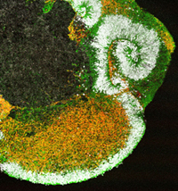
Dec 16, 2013 The characteristic wrinkled surface of the human neocortex is one the keys to the brains capacity for rational and analytic thought and language, and evolved as a means of increasing surface area to a remarkable extent. Studies of cerebral development, however, have largely been confined to model organisms, such as mouse, leaving gaps in our understanding of just this sort of human-specific feature. What is needed is a better way to study the same processes in vitro. Taisuke Kadoshima and colleagues in the Lab for Organogenesis and Neurogenesis (Yoshiki Sasai, Group Director) have now succeeded in recapitulating the human neocortical development up to the equivalent of second trimester development in an in vitro environment, building on previous work by the same lab in inducing self-organized tissue differentiation from embryonic stem cells (ESCs). This work was featured on the cover of The Proceedings of the National Academy of Sciences, in which it was published.
The Sasai group had previously shown that a 3D culture technique known as SFEBq could be used to induce steer the differentiation of human ESCs into a four-layered cortical-like structure, akin to that of a first-trimester fetus. Taking that study as their starting point, the group revisited the culture media and growth factors used in that protocol, and identified a number of opportunities for optimization, enabling even higher efficiency cortical induction. Using this modified SFEBq system, they found that nearly all ESC aggregates became positive for the telencephalic marker gene foxg1 on day 26, and rapidly began to differentiate into neuroepithelium by day 34. Following progress by a combination of live imaging and marker gene expression, the group observed the spontaneous onset of dorsocaudalventrodorsal polarity in the neuroepithelium. The dorsocaudal epithelium further began to exhibit a rolling form of morphogenesis, resulting in curvature of the inward tissue surface, evaginating to form a hemispheric, enclosed cavity, closing resembling the process of brain formation in vivo. The previous SFEBq method had made culture possible for up to around 45 days, but tweaks to the modified approach resulted in a dramatic lengthening of this time span, making it possible for the hESC-derived brain-like tissue to mature even more in vitro. By around day 70 of culture, the neuroepithelium had thickened to more than 200 µm, and showed a six-layer laminar structure corresponding to that seen in the fetal neocortex. By day 91, the tissue had deepened to 350 µm, and each layer had grown morphologically similar to that seen in a second-trimester fetus. Tracing of the movements of labeled cells over time revealed another distinctive feature of neocortical development, in which later born superficial neurons localized more superficially than older, deeper layer neurons, resembling a similar inside-out pattern seen in fetal corticogenesis. Another intriguing finding was that region of the in vitro tissue corresponding to the outer subventricular zone developed large numbers of basal progenitors akin to outer radial glia found abundantly in human. These new refinements to the SFEBq culture system will allow us to begin to more closely study important questions, such as the mechanisms underlying the spontaneous acquisition of polarity by the neuroepithelium, or the processes involved in its curvature and rolling morphogenesis, says Sasai. These are exciting times for the study of human neurodevelopment. |
|||||||
|
|||||||
|
|||||||
 |
|||||||
| Copyright (C) CENTER FOR DEVELOPMENTAL BIOLOGY All rights reserved. |