RIKEN Center for Developmental Biology
2003 Annual Report
|
|
RIKEN Center for Developmental Biology 2003 Annual Report |
Laboratory for Evolutionary Regeneration Biology
|
Its position
on an important but scientifically under-explored branch of the evolutionary
tree and its remarkable biology combine to make the planarian flatworm
a fascinating and invaluable model for basic research in fields from evolutionary
development to regenerative medicine. Kiyokazu
Isolating planarian stem cells Agata's laboratory has developed a method for identifying subsets of planarian stem cells, also called neoblasts. These somatic stem cells are the only mitotically active cells in the planarian body, making them susceptible to X-ray irradiation. Examination of cell populations shown to be vulnerable to elimination by X-rays, revealed that planarian neoblasts, like stem cells in other species, seem to exist in proliferating and resting states. Real-time PCR analysis indicates that each of these sub-populations expresses discrete sets of genes. One fraction, X1, expresses genes specific to actively cycling cells, while a second fraction, X2, lacks this expression profile and is thought to comprise stem cells in a state of quiescence. Interestingly, many X1 cells also express signal receptor molecules which are switched off in the X2 fraction, while the corresponding ligands are expressed in a variety of differentiated cells resistant to X-ray irradiation.
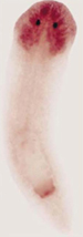
Brain regeneration by stages A planarian can re-grow
a fully functional brain within five days following the amputation of
its entire head, an extraordinary feat of self-healing that involves recapitulating
the development of the worm's entire nervous system. Researchers in the
Agata group studied this process and identified patterns of gene expression
that indicate the regeneration of the brain comprises five distinct stages.
In the first stage, at about eight hours after wound closure, a noggin-like
gene (Djnlg) is activated in
the stump prior to the formation of a blastema, a mass of
A parallel study revealed that these two genes, 1020HH and eye53, are necessary for planarians to regain their normal light avoidance behavior, known as negative phototaxis. Knockdown of these genes by RNAi had no discernible effects on brain or eye morphology, but the worms failed to respond to light stimuli in the normal manner, even after five days of regeneration, when the structure of the brain has been fully reinstated.
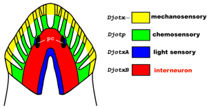
Evolution of the central nervous system The planarian is one
of the lowest forms of animal known to possess a central nervous system,
making it an apt model for the study of the evolution of this most complex
and elaborately organized biological system. Using clones of over 3,000
The Agata lab is also now participating in an international collaboration established to sequence and annotate a set of more than 10,000 planarian ESTs, which promises to provide an invaluable resource for the study of evolutionary biology, comparative genomics, and the genetics underlying the unique characteristics of these organisms. The availability of an annotated database of planarian cDNAs may provide keys to the understanding of stem cell biology, tissue plasticity and maintenance and other fundamentally important processes. |
|
Selected Publications
Agata K. Regeneration and Gene Regulation in Planarians. Curr Opin Genet Dev 13:492-6 (2003).
Mineta K, Nakazawa M, Cebria F, Ikeo K, Agata K and Gojobori T. Origin and Evolutionary Process of the CNS Elucidated by Comparative Genomics Analysis of Planarian ESTs. Proc Natl Acad Sci U S A 100:7666-71 (2003).
Nakazawa M, Cebria F, Mineta K, Ikeo K, Agata K and Gojobori T. Search for the Evolutionary Origin of a Brain: Planarian Brain Characterized by Microarray. Mol Biol Evol 20:784-91 (2003).
Agata K, Tanaka T, Kobayashi C, Kato K and Saitoh Y. Intercalary Regeneration in Planarians. Dev Dyn 226:308-16 (2003).
Saito Y, Koinuma S, Watanabe K and Agata K. Mediolateral Intercalation in Planarians Revealed by Grafting Experiments. Dev Dyn 226:334-40 (2003).
Cebria F, Kobayashi C, Umesono Y, Nakazawa M, Mineta K, Ikeo K, Gojobori T, Itoh M, Taira M, Sanchez Alvarado A and Agata K. FGFR-related gene nou-darake restricts brain tissues to the head region of planarians. Nature 419:620-4 (2002).