Crest commencement
Pax3 and Zic1 co-activate neural crest differentiation
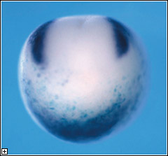
Early in vertebrate development, the foundations of the nervous system
are laid down in specific regions of the embryonic body. A sheet of epithelial
tissue rolls into a cylinder, forming the neural tube, the structure that
will give rise to the central nervous system. A migratory population of
cells called the neural crest develops slightly later, before spreading
throughout the body to create the peripheral and autonomic nervous systems,
as well as a range of other tissues including facial cartilage and bone,
the pigmented cells called melanocytes, and the adrenal medulla. Despite
the importance of the neural crest, however, the molecular signals that
function upstream in the multistep process of the specification and demarcation
of its developmental field have so far remained a mystery.
A host of regulatory molecules, including members of the BMP and Wnt
signaling families, have been implicated in this determination process,
and a pair of transcriptional factors, Foxd3 and Slug,
has been identified as definitive markers of the presumptive neural crest,
but the factors that define its exact boundaries have stayed out of reach.
In a study published in the April edition of the journal Development,
Yoshiki Sasai (Group Director, Laboratory for Organogenesis and Neurogenesis)
and colleagues reported the identification of a pair of overlapping regulatory
signals that seem to initiate the neural crest developmental program in
the African clawed frog, Xenopus laevis.
Earlier studies in the same laboratory had suggested a role for Zic-family
factors in neural crest development, and they focused on Zic1,
which is expressed in the dorsal ectodermal region of the gastrulating
embryo, the site of prospective neural development. A second molecule,
Pax3, shows a similar but distinct pattern of expression in about
the same region and embryonic stages, which led the Sasai group to narrow
their search to these candidates. Preliminary tests showed that an increase
in BMP signaling, a potent neural inhibitor, suppressed the expression
of both, while the suppression of BMP caused an expansion of their range
toward the ventral side of the embryo. Conversely, the soluble factor
Wnt caused Pax3 and the presumptive neural crest marker, Foxd3,
to be expanded beyond their normal anterior limits. They next looked at
the effects of gain of Pax3 and Zic1 function in the
developing frog, and found that both were able to trigger neural crest
differentiation, as evidenced by the expression of Foxd3 and
Slug prior to the late gastrula stage, when those markers normally
first appear, as well as in the typically non-neural ventral region. When
misexpressed singly, both Pax3 and Zic1 showed the ability
to trigger an ectopic expansion of Foxd3 and Slug in
the dorsal region, but that effect did not extend to the ventral side.
On direct injection of both Pax3 and Zic1 into the ventral
side of animal blastomeres from very early embryos, they found that the
factors in combination could indeed induce neural crest markers even in
the ventral side, indicating the potency and directness of their effect.
Sato et al followed up by studying how a loss of these molecules' function
might affect the neural crest in otherwise normal embryos by injecting
morpholino (MO) antisense oligonucleotides (a method of inhibiting the
function of specific genes by interfering with the translation of the
proteins they encode). The injection of either Pax3 or Zic1
MOs was sufficient to suppress the expression of the marker Foxd3,
while the loss of function of either of the two factors had no discernible
effects on the expression of the other, suggesting that both must be active
to achieve normal determination of the neural crest.
Animal
cap assays, which provide an in vitro model of many aspects of early Xenopus
development, helped to clarify the details of the molecular interactions
at work. Finding that Pax3 alone failed to induce Foxd3,
as it had in vivo, they began to search for the missing signals needed
to achieve that effect. When they co-injected Wnt3a (a known
factor in neural crest differentiation), they found not only that Pax3
now strongly induced Foxd3, but also that Zic1 began
to be expressed. Injection of Zic1 alone into untreated animal
caps was able to induce Foxd3, but only weakly, an effect that
was strongly complemented by co-injection with Wnt3a. Interestingly,
the inductive action of these factors acting alone could be blocked by
increasing the activity of the neural inhibitor, BMP4, but the combination
of Pax3, Zic1 and Wnt3a proved able to induce
Foxd3 robustly even in the face of an antagonistic BMP signal.
By interfering with gene function in dissociated cells, the group tested
whether this co-activity between Pax3 and Zic1 in Wnt
treated cells relied on external signals. Morpholino blockade of Zic1
in Pax3-injected and Wnt-treated single cells resulted in the
loss of Foxd3 induction, while cells exposed to all three signals
continued to express Foxd3, indicating that the Pax3, Zic1,
and Wnt3a effect is cell-autonomous. The critical role of the
endogenous Wnt cascade was shown by the loss of Foxd3 induction
when Wnt signaling was disrupted by morpholino knockdown of β-catenin,
a Wnt downstream factor.
This comprehensive and compelling set of evidence points strongly to
a modus of neural crest differentiation involving the close cooperation
of Pax3 and Zic1 in the presence of Wnt signaling in
the pre-neural embryo. That this trio of signals operates even in the
presence of inhibitory BMP signal suggests that the combination is a powerful
determinant of the prospective neural crest, and the question of exactly
how the Pax3-Zic1 partnership overrides BMP on the molecular level represents
an intriguing subject for further study.