| RIKEN Center for Developmental Biology (CDB) 2-2-3 Minatojima minamimachi, Chuo-ku, Kobe 650-0047, Japan |
Now, in an article published in the journal Development, Koji Tanabe and colleagues in the Laboratory for Cell Adhesion and Tissue Patterning (Masatoshi Takeichi; Group Director) report the use of a new gene transfer system that enabled them to study the function of cadherins in horizontal cells in the embryonic retina in chicken. These cells, which receive input from photoreceptors and modulate intercellular feedback in the retinal network, form elaborate dendritic fields that enable them to make contacts with multiple surrounding neurons. Tanabe, who later moved to the RIKEN Frontier Research Program Nakagawa Research Initiative Unit (Shinichi Nakagawa; Unit Leader), found that in neurons in which cadherin function had been lost, the size of these fields was smaller and that the individual dendrites frequently failed to connect properly with photoreceptor cells.
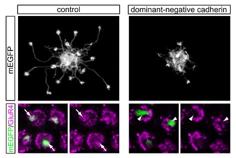
The ability to observe horizontal cells as they developed in their natural habitat enabled the group to watch as the initially indistinguishable cells gradually transformed, extending dendrites that went on to form synaptic connections in patterns specific to their morphological type. Type I dendrites were found to project to both principal and accessory populations of double cone photoreceptors; dendrites from Type III cells, however, terminated only in accessory cones. The picture of Type II horizontal cells was less clear, as there are no molecular markers capable of distinguishing their targets at the resolution needed. After verifying that horizontal cells express the cadherin family member N-cadherin in vivo, Tanabe used a plasmid vector to overexpress a dominant-negative version of this molecule together with mEGFP in horizontal cells, thereby blocking cadherin function. The cadherin-inhibited cells labeled by mEGFP diversified into the three horizontal cell morphologies, but in all three types, the dendritic fields were smaller than normal.Axon outgrowth, in contrast, was unaffected. Looking next at the level of individual dendrites, Tanabe found that although dendrites appeared to home in to their appropriate targets, the morphologies of the dendritic terminals were severely affected. Moreover,Tanabe showed thatcadherin regulates synapseformationbyobservingthat, eveninlessmorphologicallyaffecteddendritic terminals,cadherinblockageperturbedtheaccumulationof the synapticmarkerGluR4. Given that the cadherin-negative horizontal cells showed defects in both the global organization of the dendritic and the local ability to form functional synapses, Tanabe et al. suggest that cadherins may be involved in two distinct stages of dendritic morphogenesis; first as the dendrites elongate, then again later as they form synaptic contacts. This represents an important in vivo demonstration of cell adhesion machinery in synapses, and a new stride forward toward a better understanding of how the neural network forms from assemblies of initially discrete cells. |
|||||
|
|||||
 |
| Copyright (C) CENTER FOR DEVELOPMENTAL BIOLOGY All rights reserved. |