Labs

Cell Adhesion and Tissue Patterning

Team Leader
Masatoshi Takeichi (Ph.D.)

Animal cells organize into tissues with complex architecture. Our lab is exploring the molecular mechanisms by which individual cells assemble into a tissue-specific multicellular structure, such as epithelial sheets and neural networks. We are also interested in the molecular basis of how tissue architecture is disrupted during carcinogenesis, a process that is thought to accelerate the metastasis of cancer cells. For these studies, we are focusing on the roles played by cell-cell adhesion and recognition molecules, the cadherin family of adhesion molecules in particular, as these are known to be indispensable for tissue construction. Our current studies are divided into three categories:

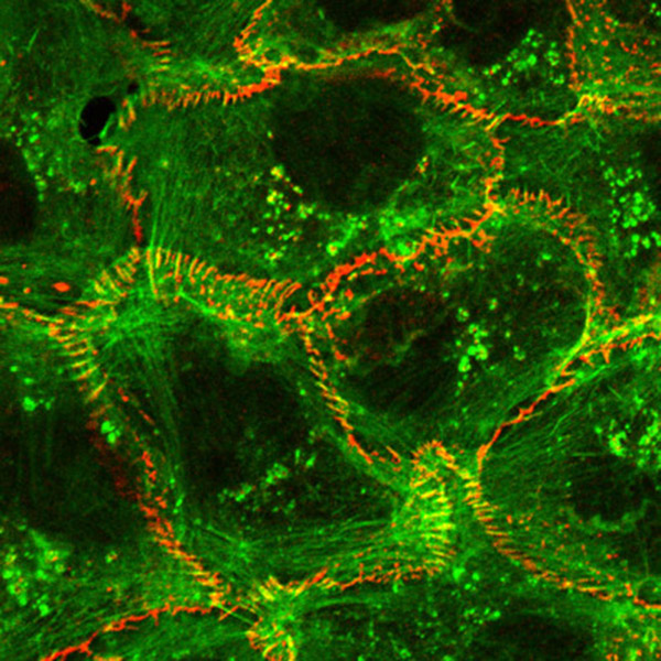
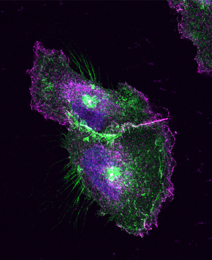
1) Cell-cell adhesion is a dynamic process, and this nature of cell-cell adhesion is implicated in various cell behaviors, such as contact-dependent regulation of cell movement and cancer metastasis. A growing body of evidence suggests that cadherins cooperate with cytoskeletal and/or motility machineries, such as actin regulators, non-muscle myosins, and Rho GTPases, in modulating cell assembly. We are therefore studying the molecular mechanisms underlying the crosstalk between cadherins and such cytoskeletal systems, and their roles in epithelial junction formation.

2) A second area of interest to our lab is to gain a better understanding of how the cell-cell adhesion machinery contributes to animal morphogenesis. Using mouse embryos, we are analyzing the roles of cadherins and associated proteins in various morphogenetic processes, including neural crest migration. We are also investigating the roles of members of the cadherin superfamily known as protocadherins, deficiencies of which have been implicated in human brain disorders. Through these studies, we expect to gain deeper mechanistic insights into the ways by which cells build the elaborate structures of the animal body.

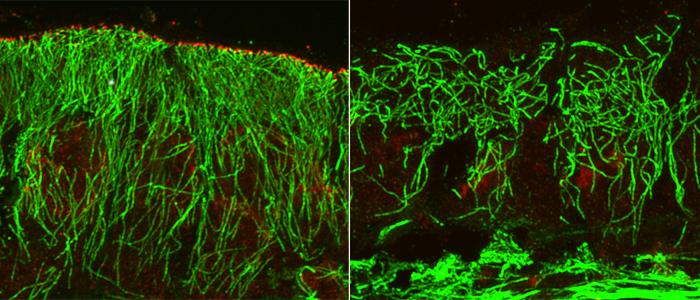
3) In addition, we have been analyzing the functions of microtubule minus end-associated proteins, Nezha/CAMSAPs. These proteins regulate microtubule assembly patterns, centrosomal function, and organelle positioning. We are exploring the roles of these molecules in cellular morphogenesis, such as polarized epithelial formation and axon growth, with the aim of uncovering novel functions of non-centrosomal microtubules.

News List
2018.3.19
Induced tension restores apical junctions in carcinoma cells
2018.3.15
Catenins guide cell migration
2018.3.14
Loss of PCDH19 leads to differential behavior between sexes
2017.2.1
DAAM1 stabilizes adhesion between lateral membranes of epithelial cells
2016.2.26
Mechanism orienting microtubules in epithelial cells
2015.2.5
Strategy of bending by giant cadherins
2014.12.18
Protocadherin mediates collective axon extension of neurons
2013.10.1
New roles for microtubules in actin regulation
2012.12.3
CAMSAPs regulate microtubule dynamics in epithelial cells
2012.11.12
N-cadherin keeps cornea pumping
2012.9.19
CDB Director Masatoshi Takeichi named citation laureate
2012.5.28
Celsr1 bridges key processes in formation of neural tube
2011.10.12
EPLIN responds to force
2011.7.10
Dual role for AIR-1 in mitotic spindle assembly
2011.6.22
Willin and Par3 work together in apical constriction
2009.7.21
Fat and Daschous cadherins work at the apical membrane in cerebral cortical cells
2009.7.14
Synapses stay true: Selective neural connectivity in vitro
2008.12.9
Tracks to the junction: Microtubules tethered to zonula adherens
2008.8.6
Pairing up and settling down
2008.4.25
Shroom3 recruits ROCKs to the apical junctions
2007.12.17
A mystery of the cadherin-actin interaction resolved
2007.8.30
Protocadherin in neural pathways
2007.5.8
CDB Director Masatoshi Takeichi elected to National Academy of Sciences
2007.4.6
Cold proof: Cadherin-8 in neural circuitry and cold sensation
2007.2.19
Catenin cleft: Calpain-mediated cleavage of β-catenin in novel signaling pathway
2006.12.25
Cadherins go with the flow
2006.10.31
Keeping an eye on cadherin function in retinal synaptogenesis in vivo
2006.10.21
Tuba joins the band: A Cdc42-specific activator regulates epithelial cell-cell junctions
2006.7.29
Neurite sorting by nectins
2006.5.12
Binding to build the brain
2005.9.12
CDB Director Elected President of ISDB
2005.5.20
2b or not 2b a stem cell
2005.1.13
Masatoshi Takeichi Awarded 2005 Japan Prize
2004.12.14
France, Japan Recognize CDB Director’s Work
2004.9.13
Interview with CDB Director, Masatoshi Takeichi
2004.5.26
Fat binds the “bones” of the cytoskeleton
2004.5.12
CDB Director elected to American Academy of Arts and Sciences
2004.4.15
Dendritic spine dynamics
Select References

Ito S, et.al. Induced cortical tension restores functional junctions in adhesion-defective carcinoma cells. Nature Communications 8, 1834 (2017) doi:10.1038/s41467-017-01945-y

Vassilev V, Platek A, et.al. Catenins Steer Cell Migration via Stabilization of Front-Rear Polarity. Dev Cell. 2017 Nov 20. doi: 10.1016/j.devcel.2017.10.014.

S Hayashi, et.al. Loss of X-linked Protocadherin-19 differentially affects the behavior of heterozygous female and hemizygous male mice. Scientific Reports. 2017 July 19. doi: 10.1038/s41598-017-06374-x

Nishimura T, et al. DAAM1 stabilizes epithelial junctions by restraining WAVE complex-dependent lateral membrane motility J Cell Biol 215, 559–573 (2016). doi:10.1083/jcb.201603107

Toya M, et al. CAMSAP3 orients the apical-to-basal polarity of microtubule arrays in epithelial cells. Proc Natl Acad Sci U S A 113, 332–337 (2016). doi:10.1073/pnas.1520638113

Tsukasaki Y, et al. Giant cadherins Fat and Dachsous self-bend to organize properly spaced intercellular junctions. Proc Natl Acad Sci U S A 111, 1601–1606 (2014). doi: 10.1073/pnas.1418990111

Hayashi S, et al. Protocadherin-17 mediates collective axon extension by recruiting actin regulator complexes to interaxonal contacts. Dev Cell 30, 673–687 (2014). doi:10.1016/j.devcel.2014.07.015

Takeichi M. Dynamic contacts: rearranging adherens junctions to drive epithelial remodelling. Nat Rev Mol Cell Biol 15, 397–410 (2014). doi:10.1038/nrm38025

Tanaka N, et al. Nezha/CAMSAP3 and CAMSAP2 cooperate in epithelial-specific organization of noncentrosomal microtubules. Proc Natl Acad Sci U S A 109, 20029–20034 (2012). doi: 10.1073/pnas.1218017109

Q:What made you want to do your current job? (or triggered your interest in science)

A:…

Q:What would you ultimately like to discover through your research?

A:…

Lab Homepage

takeichi[at]cdb.riken.jp

Recruit

Double-immunostaining for F-actin (green) and Kusabira Orange-tagged E-cadherin (red) introduced into A431D cells. In these cells, E-cadherin dynamically moves along cortical actin filaments, resulting in the unique distributions shown here.
A pair of U251 cells in contact with one another, triple-immunostained for Protocadherin17-EGFP (green), the WAVE complex protein Abi-1 (magenta) and DNA (blue). Protocadherin17 recruits the WAVE complex to cell-cell contact sites, and convert them into a motile structure. Cell edges with this structure actively move, contributing to collective cell migration.
Microtubules (green) and CAMSAP3 (red) in intestinal epithelial cells. CAMSAP3 localizes at the apical cortex of the cells, and tether microtubules to this site through its binding to their minus ends. When CAMSAP3 is knocked out, the longitudinal arrays of microtubules are disrupted.
PAGE TOP