| RIKEN Center for Developmental Biology (CDB) 2-2-3 Minatojima minamimachi, Chuo-ku, Kobe 650-0047, Japan |
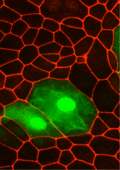
June 22, 2011 Epithelial cells often take the shape of truncated wedges, with an apical surface narrower than the basal face. This form of cellular polarization plays a central role in morphogenetic processes in which sheets of homogeneous tissue bend, fold over, or roll up due to minute changes in the geometry of their component cells. The primary connections between cells in such epithelial sheets are known as apical junctional complexes (AJCs), which both bind cells of like type to their neighbors and engage with the cytoskeleton elements called actomyosin cables to modify and reinforce cell shapes and behaviors. The contractility of such cables is under the control of a family of kinases known as ROCK proteins, which form complexes near AJCs and enhance actomyosin contraction through phosphorylation of myosin light chain. But how ROCK interacts with factors involved in cell polarity remains something of a mystery.
Takashi Ishiuchi in the Laboratory for Cell Adhesion and Tissue Patterning (Masatoshi Takeichi, Group Director) has begun to tie the threads together with the discovery that a previously enigmatic protein called Willin works cooperatively with the known polarity protein Par3 to phosphorylate ROCK, a critical trigger of apical constriction. Published in Nature Cell Biology, this new work shed light on the complex regulation of this important cellular morphogenetic process. The study began with an examination of Willins localization, which revealed that it clusters near the apical junction. A subsequent immunoprecipitation assay turned up an interaction with Par3, catching the groups attention, due to Par proteins involvement in cell polarization. They tested whether expression of Willin would affect Par3 localization, and found that while this had no effect, it did increase the accumulation of Par3 partner aPKC at the cell membrane. A second round of immunoprecipitation showed that Willin associates with aPKC as well, which deletion mutant studies revealed to be dependent on a region in the Willin protein known as JFR. As Willin expression had no measurable effect on Par3 levels, Ishiuchi tried the converse experiment and showed that Par3 is not required for Willins upregulation of aPKC. Willin was also not required for the Par3-aPKC association, indicating that while Willin could interact with both of these proteins, it appeared to do so by independent mechanisms. Curious about Willins true role, the group knocked down the function of both Willin and Par3 in cells, and found that this resulted in tightening of the apical junctional complex, secondary to a dramatic reduction in the localization of aPKC near the membrane. It appeared that aPKC was the critical downstream factor for limiting apical constriction. This was confirmed by experiments using a membrane-targeted aPKC construct, which was able to rescue excessive actomyosin contractility. But how does aPKC achieve this effect? Ishiuchi had observed that ROCK inhibition abrogated apical constriction, so he turned his focus to possible interactions between the two factors, with an intuition that this activity might rely on phosphorylating activity by aPKC. On treating ROCK1 with a phosphatase, he found that this caused a change in its molecular mass, suggesting that the protein is indeed phosphorylated in vivo, which the group was then able to verify using a system to detect phosphorylation by band-shift. Analysis of the distribution of wildtype and phosphorylated ROCK proteins revealed that while wildtype ROCKs tended to aggregate at the apical junction, when phosphorylated they remained in the cytoplasm. The Par3-aPKC complex is known to regulate the apico-basal polarity of cells, and Willin is thought to be a relative of the Drosophila protein Expanded, a component of the Hippo pathway for growth control, says Takeichi. It will be interesting to work out how the ability of these proteins to regulate actomyosin contraction at AJCs is related to their other physiological functions in future studies. | |||||
|
|||||
 |
| Copyright (C) CENTER FOR DEVELOPMENTAL BIOLOGY All rights reserved. |