| RIKEN Center for Developmental Biology (CDB) 2-2-3 Minatojima minamimachi, Chuo-ku, Kobe 650-0047, Japan |
July 14, 2009 The brain is a bramble, with thorny branches of neuronal projections intertwining into a dense network. But a closer look reveals an order to its seemingly thicket-like structure, in which axons from one neuron connect with dendrites from specific partners. In cultured neurons, however, these relationships become blurred, and synapses have been shown to form between mismatched neurons, raising questions about the mechanisms that control neuronal recognition in vitro.
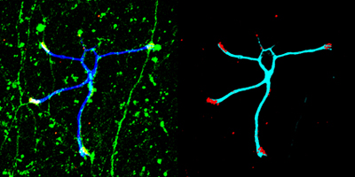
Shoko Ito of the Laboratory for Cell Adhesion and Tissue Patterning (Masatoshi Takeichi; Group Director) has reexamined this notion of in vitro neuronal promiscuity and found that, in contrast to previous allegations, at least some dendrites remain true to their partners. In studies of synapse formation using cerebellar granule cell dendrites, Ito showed that while these may come into contact with axons of all types in cell culture, they synapse only with mossy fiber axons, just as they do in vivo. This work is published in the Proceedings of the National Academy of Sciences. The cerebellum features two characteristic neuronal cells types: the huge and hugely intricate Purkinje cells, which form synapses with climbing fibers from a medullary region called the inferior olivary nucleus, and granule cells which synapse with mossy fiber axons from a different region known as the pontine nuclei. Ito began with a neuronal co-culture system in which granule cells were grown in the presence of climbing fibers and hippocampal neurons, neither of which would they ordinarily form synapses with in the brain. These explants projected axons as normal, and the granule cells extended dendrites, but although they in some cases the neurites came into close contact with each other, synapses of the type seen in vivo failed to form. Only when granule cells were cultured with their typical binding partners, mossy fiber axons from the pontine, did they form synapses which are functionally normal and morphologically identical to those seen in vivo, as indicated by the expression of synaptic markers. Axons from the inferior olivary and hippocampal cultures also apparently formed synapses, but on closer examination these showed their arrangement was abnormal, and they showed impaired release of synaptic vesicles, which are responsible for the cycling of neurotransmitters. This new work clearly shows that the selective synapse formation known to occur in vivo is replicable in cultured neurons as well. The mechanisms by which such selection takes place, however, remain unknown and must await further studies now that this fundamental principle has been shown to be conserved in vitro. |
|||||
|
|||||
 |
| Copyright (C) CENTER FOR DEVELOPMENTAL BIOLOGY All rights reserved. |