New work by Shinsuke Nakao and others in the Laboratory for Cell Adhesion and Tissue Patterning (Masatoshi Takeichi; Group Director) now adds another piece to this puzzle with the revelation that another cadherin known as OL-protocadherin is involved in recruiting cytoskeletal regulators to cell-cell contact sites, a previously unknown means of modulating cell migration.
OL-protocadherin (OL-pc) shows a similar like-binds-like mode of interaction to that seen in other cadherins, although its homotypic bonds are comparatively weaker. Previous work from the Takeichi lab had shown that rather than acting as a straightforward adhesion molecule, OL-pc might instead function in other cellular processes – neurons in mice lacking OL-pc, for example, showed defects in axon extension.
This novel role prompted the group to look for interactions with other molecules, using a technique known as a pull-down assay, in which the OL-pc protein was used as “bait” to survey for other proteins that bind with it. Their search for binding partners turned up a protein named Nap1 that is known to associate with a second factor called WAVE1, which functions in the assembly of the actin cytoskeleton protein into forms necessary for cell migration.
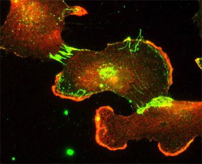
After identifying the specific molecular region at which Nap1 binds to OL-pc, Nakao et al. next investigated its cell biological function by transfecting OL-pc into a line of cells that shows high motility when cultured at low densities. Typical of other cadherins, OL-pc tends to accumulate in regions of contact between cells, while Nap1 and WAVE1 are concentrated in lamellipodia. In cells transfected with exogenous OL-pc, both Nap1 and WAVE1 became detectable at cell-cell contacts as well, phenotypes that were not seen when OL-pc lacking the Nap1-binding region was used.
As Nap1 and WAVE1 are known to regulate cell movement, Nakao were curious about a possible role for OL-pc in this process as well. In cells moving freely at low culture densities, OL-pc misexpression had no apparent effect when observed using time-lapse microscopy. But when the cells were grown in more crowded quarters, enabling the formation of stabler cell-cell contacts, OL-pc-expressing cultures showed higher motility than their untransfected counterparts, with the cells moving in a jumpy, uncoordinated fashion. Tests in which Nap1 and WAVE1 were knocked down using RNAi revealed that OL-pc’s function relied at least in part on the function of these actin regulators.
Interestingly, cells lacking N-cadherin, a classic cadherin expressed in neurons, mimicked the OL-pc misexpression phenotype, which, combined with observations of altered organization of adherens junctions in transfected cells, suggests that the OL-pc-Nap1-WAVE complex acts by remodeling actin assembly at cell-cell contact sites, causing changes in the structure of the cell-cell junction. The group speculates that this may be the basis for the increase in cell motility, in contact inhibition mediated by cadherin adhesion is weakened due to the alteration of cell-cell junction structure. |