| RIKEN Center for Developmental Biology (CDB) 2-2-3 Minatojima minamimachi, Chuo-ku, Kobe 650-0047, Japan |
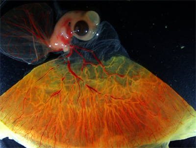
June 30, 2008 – In chickens, as in other vertebrates, the emergence of the blood system takes place in two waves, a first that originates in extraembryonic tissue, and a second within the developing embryo itself. The nature of this early hematopoiesis is of great potential interest to scientists and clinicians alike, but the small size of the embryo in most popular model organisms makes it difficult to obtain enough material to conduct comprehensive studies. The comparatively large size of the chicken embryo at the transition from primitive (extraembryonic) to definitive hematopoiesis in the embryo proper makes it a good platform for investigating the changes that take place during this critical stage in the development of the blood system.
Brendan McIntyre and others in the Laboratory for Early Embryogenesis (Guojun Sheng; Team Leader) took advantage of this plentiful supply to examine gene expression profiles of blood during the period between embryonic days 4 and 6, when the specification of definitive hematopoietic lineages occurs. Through microarray data analysis, he found that circulating non-red blood cells include a full complement of blood progenitors and stem cells, meaning that this population may serve as a useful alternative to embryonic stem cells and perinatal blood for scientists interested in embryonic hematopoiesis. The team began by taking samples from chicken embryos at days E4 and E6, and centrifuging them to sort the heavier red cells from the other lighter components of the blood system. Additional cell sorting by FACS and classical staining techniques enabled them to isolate a population of cells from which all red blood had been excluded. RT-PCR, a technique for amplifying gene transcripts, showed that the non-red blood population expressed the hematopoietic stem cell (HSC) marker CD34, while the red blood aliquot did not, indicating that the non-red sample represented the sole source of blood stem cells in the embryo. McIntyre et al. next used Affymetrix gene chips to compare levels of gene expression, and identified a number of candidates whose expression was higher in the non-red blood cells (but significantly was not elevated in the heart). Their analysis revealed that numerous genes associated with hematopoietic stem cells, including those used in cell signaling, cell-matrix adhesion and communication between cells at gap junctions. In addition to these HSC genes, the team found markers of the myeloid and lymphoid lineages, providing the first evidence in chick of lymphoid differentiation at this early stage of development. The search also yielded 6 functionally uncharacterized genes, which it is hoped will provide new insights into HSC function and differentiation on further study.
|
|||||
|
|||||
|
|

| Copyright (C) CENTER FOR DEVELOPMENTAL BIOLOGY All rights reserved. |