

|
 |
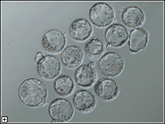
Aft agleyA prescription for reprogramming errors in nuclear transferMammalian cloning remains a young field whose first fruits, such as Dolly the sheep and Cumulina the cloned mouse, are still less than a decade old. When Teruhiko Wakayama (Team Leader; Laboratory for Genomic Reprogramming) and colleagues at his former lab under Ryuzo Yanagimachi in the University of Hawaii successfully created the first cloned mouse by transferr-ing the nucleus of an ovarian cumulus cell to an unfertilized egg, it was heralded as a major achievement in the face of the low efficiencies that dog the procedure even to this day. Since that time, researchers have tried altering the timing of the nuclear transfer (NT), tested an array of methods to activate the oocyte into receptivity, and experimented with a whole range of differentiated cell types as nuclear donors, but to little effect; the success rate of mouse cloning attempts to produce live offspring has languished at only about 2%. A straightforward new tweak developed by the Wakayama lab, published in the 9 December 2005 edition of Biochemical and Biophysical Research Communications, presented the first significant increase in mouse cloning efficiency in recent years. This step forward was achieved by researchers who treated the NT zygotes with the histone deacetylase inhibitor, trichostatin A (TSA), thereby boosting efficiency to an unprecedented 6%. The reasons for the difficulties in the cloning of mammals have been
proposed to stem back to incomplete or incorrect reprogramming of the
nucleus following transfer into the oocyte. The chromosomes of differentiated
cells carry molecular markers that specify which genes are to be expressed
and which shut down, helping to specify the patterns of gene expression,
and so, the form and function of such cells. In natural fertilization,
the information sets contained with nuclei of the sperm and egg are reprogrammed
as the two cells fuse, enabling the ontogeny of a unique new individual.
But when a differentiated nucleus is introduced directly into an oocyte
as is done in cloning attempts, this reprogramming frequently appears
to go awry. The molecular signatures involved are written in a script
of methyl groups that adorn genes directly and the acetylation of histone
complexes that package the DNA into tightly coiled bundles, but it is
a mystery how the oocyte opens up this intricate code for revision.
The positive effects of TSA treatment translated into higher live births as well, with 6% of all attempts leading to the birth of pups (against only about 1% for control). These animals were found to be healthy and normal in all respects, save for the enlarged placenta that typifies all cloned mice, and importantly, do not exhibit the obesity and shortened life expectancy seen in some other mouse clones in the past. Interestingly, when the donor nucleus was taken from an ES rather than a somatic cell, TSA had the opposite effect, and no clones developed to term. ES cells are in themselves excellent nuclear donors, as their DNA methylation is naturally low; unmodified cloning by ES cell nuclear transfer generally sees 2~6% success in producing live pups. The picture presented by these findings is that methylation may be a determinant of success that is highly dosage-sensitive. TSA appears to be a positive addition to the technique for establishing ntES cell lines, which are embryonic stem cell lines derived from blastocysts created by nuclear transfer. TSA treatment increased efficiency by two or threefold, and the ntES cells so created were found to express all of the molecular markers characteristic of ES cells, including Oct3/4 and Nanog. Cloning from somatic cells and the derivation of ES cells from nuclear transfer embryos represent extremely promising technologies, with applications ranging from the preservation of endangered species to the generation of cell populations for clinical use free of the risk of immune rejection. For those reasons alone, this advance in the efficiency of these procedures is of unmistakable impact. The underlying evidence suggesting a critical role for histone deacetylase inhibitors in reprogramming is also intriguing for the fundamental biological insights it provides. A more detailed examination of the precise means by which TSA augments the mechanisms of reprogramming awaits. |
|
Kishigami S, Mizutani E, Ohta H, Hikichi T, Thuan N V, Wakayama S, Bui
H T and Wakayama T. Reprinted from Biochemical and Biophysical Research Communications 340, 183-189 (2005) with permission from Elsevier |