Innermost secrets of inner ear induction
The ear's architecture is elaborate, with the three distinct components
of outer, middle and inner ear coordinating to enable the senses of hearing,
motion and balance. These otic subunits arise via separate genetic programs,
the foundations of which are laid down very early in the embryo's development.
The inner ear, which grows to become home to populations of signal-transducing
hair cells used in audition and maintaining equilibrium, is a marvelously
intricate and multifunctional structure. It derives from a small patch
of embryonic tissue known as the otic placode, a thickened disc that appears
in an ectodermal region that would otherwise be destined to become skin.
Studies
in chicken and mouse have shown that the specification of this placode
is the outcome of a pattern of tissue interactions between tissues from
at least two of the three embryonic germ layers, mesoderm and ectoderm.
This previous work demonstrated that, in both species, the adjacent regions
of head mesoderm and neural ectoderm (or caudal hindbrain) must communicate
and work together to achieve complete development of the otocyst. In the
March 2005 issue of the journal Genes and Development, Raj Ladher
(Team Leader, Laboratory for Sensory Development) and colleagues at the
University of Utah have now determined a role for signals from the third
germ layer, endoderm, in the initiation of the inner ear.
The study of ear development reveals great diversity in molecular agents
across taxa, although most of that variety is confined to the FGF family
of secreted proteins and receptors. In the chicken embryo, signaling by
FGF19 from the cranial mesoderm induces the expression of WNT8c and FGF3
in the neural ectoderm, which then cooperatively induce the otic placode
to form in a nearby non-neural ectodermal region. The mouse achieves the
same ends by different means, using mesodermal FGF10 and FGF3 in the hindbrain
to instruct the placode to form. Zebrafish embryos, meanwhile, take a
third route, utilizing FGF3 and FGF8 to engage and regulate otic induction.
This last example sparked Ladher's interest, as there had been no reports
of Fgf8 expression in the parotic regions of either chicken or
mouse, and, with his colleagues in Utah, he began to investigate the possible
involvement of FGF8 in the induction of the ear in these species.
Tissue ablation studies in chick showed that endoderm makes a contribution
to the initiation of otic development, which subsequent investigations
indicated was due to its induction of mesodermal Fgf19. Looking in the
subjacent endoderm at developmental stages corresponding to the start
of the Fgf19 expression, Ladher found that Fgf8 is indeed expressed
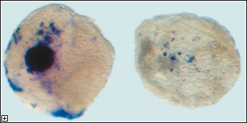
in patterns suggesting a role in otic induction. The team found that exogenous
FGF8 was sufficient to induce mesodermal Fgf19, then demonstrated
its necessity by inhibiting Fgf8 expression in vivo using RNAi,
which resulted in the downregulation of Fgf19 and failure of
otic placode development.
In parallel studies in mouse, Fgf8 was found to be expressed
in a pattern consistent with a role in the development of the ear, with
transcripts detected in relevant sites and at time-points synchronous
to otic placode induction. Gene expression patterns indicated a possible
overlap in function between Fgf8 and another FGF family member, Fgf3.
Embryos entirely lacking Fgf8 die prior to the initiation of
ear development, so the team constructed a model, in which Fgf8
expression is dramatically reduced and Fgf3 absent, to test the effect
on otogenesis. They found that, at these low levels, Fgf8 was
unable to support ear development in the absence of Fgf3. This
effect, however, did not translate to abnormalities in hindbrain development
(which contrasts with the case for zebrafish, in which Fgf3 and
Fgf8 also function redundantly), suggesting that in mouse the
ear and the hindbrain are determined by different genetic routines. Examining
the Fgf3/8 double mutants more closely, the team saw that the
phenotypes resembled those of Fgf10 knockouts, a similarity which
was substantiated at the molecular level by in situ hybridization studies
showing clear reductions in Fgf10 expression in the double mutant
mesenchyme.
"What's interesting about these results is that they show the involvement
of signals from all three germ layers in inducing the ear," says
Ladher. "In both chick and mouse, endodermal FGF8 seems to be working
as a molecular cue ball, setting off different combinations of serial
and parallel interactions in the overlying mesoderm and ectoderm that
ultimately result in the specification of the otic placode."