![]()
This movie shows time-lapse images of early Drosophila embryonic development. The embryo is labeled with membrane-GFP (Resille-GFP, green) and H2Av-RFP (magenta) and optically sectioned using two-photon microscopy to view at the mid-sagittal focal plane. Initially, the embryo forms a single layer of cells all around the entire embryonic cortex. This process is called “cellularization”. Subsequently, several distinct regions of cells undergo cell shape changes to initiate gastrulation morphogenetic movements including ventral furrow formation, posterior midgut invagination, cephalic furrow formation and dorsal fold formation. The trunk of the embryo eventually curves into a U-shape during germband extension. Scale bar is 10 μm. |
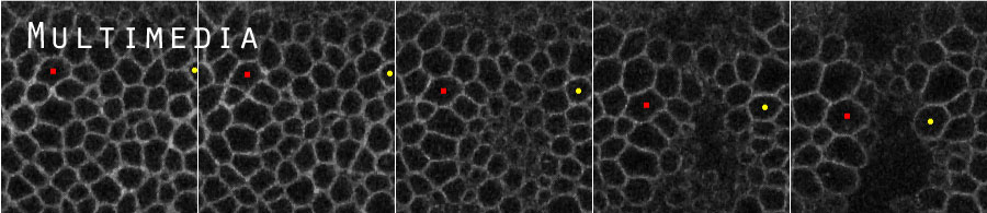
This movie shows time-lapse images of dorsal fold formation. The embryo is labeled with E-Cadherin-GFP (green) and membrane-mCherry (Gap43-mCherry, magenta). The dorsal fold region is viewed at the mid-sagittal focal plane using two-photon microscopy. Note that adherens junctions (visualized by E-Cadherin-GFP) shift basally in the initiating cells prior to cell shape changes and tissue movement. Although the initiating process is similar between the two dorsal folds, the eventual anterior fold (on the left) is shallow, while the posterior fold (on the right) undergoes extensive invagination to form a deep epithelial fold. Scale bar is 10 μm. |
This movie is a composite of four movies, each of which represents a distinct genetic condition. In contrast to the wild-type embryo in which adherens junctions become differentially positioned along the apical-basal axis of cells across the epithelium to organize the folding, embryos of par-1 or bazooka RNAi knockdown display uniform positioning of junctions laterally or apically and the folding is blocked. Conversely, overexpression of bazooka raises the ratio of Bazooka and Par-1 and promotes ectopic junctional shifts and formation of ectopic epithelial folds. |
This movie demonstrates the results of EDGE4D image processing, 3D rendering of cell shapes and cell tracking during epithelial folding. The original Z stack is produced using two-photon microscopy by imaging an embryo that expresses a membrane marker (Resille-GFP, green) and a nuclear marker (H2Av-RFP, magenta). Each reconstructed and tracked cell is rendered in 3D with a distinct color. |