
News and Announcements from the CDB
The chick has long been a cherished model for studying development, owing to the ease with which the embryo can be accessed and manipulated inside the eggshell. However, much of its first day of development following fertilization, which occurs inside the hen before the egg is laid (oviposition), remains shrouded in mystery. Studies of fish and mouse embryos show that initial cleavage processes immediately after fertilization heavily influence cell fate decisions and morphogenesis at later stages, but whether similar developmental processes unfold in avian embryos in the early stages before egg-laying has not been closely examined before.
Now a new study by technical staff Hiroki Nagai of the Laboratory for Early Embryogenesis (Guojun Sheng, Senior Investigator) along with other collaborators, including team leader Shigenobu Yonemura of the Ultrastructural Research Team at the RIKEN Center for Life Science Technologies (CLST), closes in on those early developmental processes in avian embryos that take place after fertilization but before the egg is laid. Using both light and scanning electron microscopy (SEM), they carried out detailed observations of the developmental patterning of pre-oviposition chick embryos, providing the first look at early embryonic development that occurs before egg-laying. Their findings, published in Development, reveal several surprising similarities in early developmental patterning among birds and other vertebrate species.
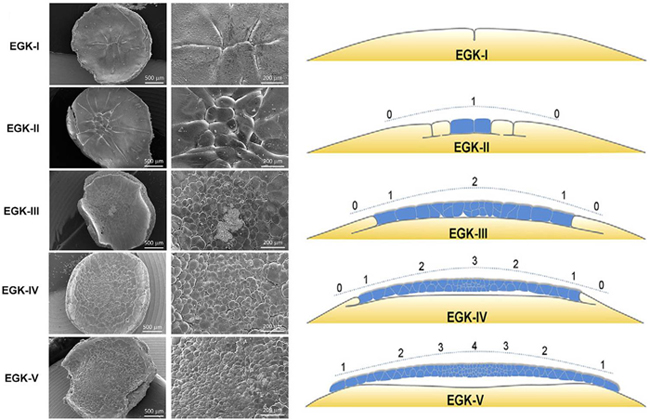
In the 1970s, scientists Eyal-Giladi and Kochav created a developmental staging system for chick embryos (EGK stages), which classifies early embryonic stages based on morphological characteristics observed under the light microscope; the staging system divides developmental processes between post-fertilization to pre-primitive streak formation into 14 stages, from EGK-I to -XIV, with EGK-I to -X covering early embryonic stages before oviposition. In the 1990s, with the help of electron microscopy, Etches and colleagues were able to observe ultrafine structures of the later pre-ovipositional EGK stages (EGK-IV onward) in chick embryo. For the current study by Nagai and colleagues, they examined the morphological characteristics of the early pre-ovipositional EGK stages (EGK-I to -V: post-fertilization to 8 hours) in detail using both light and electron microscopy, retrieving the fertilized eggs from the hens with a non-invasive, abdominal massage method.
During EGK-I, several cleavage furrows appear on the surface of the yolk on the animal pole side, forming multinucleate cells. At EGK-II, cells of varying sizes that have completed cellularization (compartmentation) are found near the center, and by EGK-III, these cells begin a series of rapid divisions and a space between cellularized cells and yolk cells, called the subgerminal cavity, emerges.
The team then focused on two important events that occurs during early developmental stages to look for possible similarities with other vertebrate species. One event was zygotic gene activation (ZGA), when the embryo, which is initially dependent on maternal transcription products accumulated in the egg for development, switches over to its own developmental regulatory system by activating transcription of its own genes. ZGA is thought to be closely linked to epigenetic reprogramming mechanisms in the cells, but when and how this occurs has not been well studied in avian species. Upon examining the chick embryos, they discovered that ZGA takes place between late EGK-II and early EGK-III (64- to 128-cell stages), which is similar to zebrafish embryos in which ZGA occurs at the 128-cell stage. That ZGA occurs at similar developmental timing in chick and zebrafish, which both have yolk-rich eggs, suggest there is a common embryonic molecular mechanism at work.
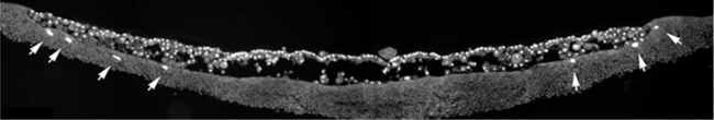
The second event the group focused on was one specific to unevenly distributed yolk-rich eggs such as chick and zebrafish, the formation of the yolk syncytial layer (YSL). Research in zebrafish have revealed that the YSL forms during the cleavage stage near the boundary of where the embryo (blastoderm) contacts the yolk and contributes to the dynamic collective migration of blastoderm cells; whether a similar YSL forms in avian embryos has remained unclear. Nagai et al. carefully examined the pre- and post-oviposition chick embryos and found a YSL was present between stages EGK-V to -XI. Analyses of other avian species, such as finches and quails, also showed formation of YSL at similar developmental timings as in the chick. These findings indicate that YSL formation is a common developmental phenomenon among birds and fishes.
“Several features of early embryonic development in chick closely resemble what is seen in zebrafish embryos,” says Sheng. “It will be interesting to determine whether the features conserved across species are a result of convergent evolution or of evolutionary constraints.”
| Link to article |
|---|
![CDB [RIKEN CENTER FOR DEVELOPMENTAL BIOLOGY]](http://www.cdb.riken.jp/en/wp-content/themes/cdb_en/images/common/fLogo2.png)
2-2-3 Minatojima-minamimachi, Chuou-ku, Kobe 650-0047, Japan
TEL : +81-78-306-0111
FAX : +81-78-306-0101
E-mail : cdb[at]cdb.riken.jp

