
News and Announcements from the CDB
Following tooth loss, dental treatments using artificial materials such as dentures, dental bridges and implants are used to replace the tooth, and however these methods do not restore full physiological function of the teeth. Other tooth replacement therapies such tooth transplantation of wisdom tooth and transplantation of autologous or bioengineered tooth germs, a group of cells in early development that give rise to teeth, into regions of tooth loss, have recently been developed and shown to fully restore physiological tooth function. As humans have a limited number of tooth germs, securing biological sources that do not cause immunological rejection remains a critical issue, and thus the development of a technique to increase tooth germ numbers is heavily anticipated.
A recent research collaboration between CDB’s Takashi Tsuji, team leader of the Laboratory for Organ Regeneration, and graduate student Naomi Yamamoto and others in Prof. Keiji Moriyama’s laboratory at the Tokyo Medical and Dental University has led to the development of a unique approach for tooth regeneration, using a mouse model. They split a single tooth germ into two using mechanical force, which could subsequently be transplanted into the oral cavity and develop into two fully functional teeth. Their approach and findings are published in the online journal, Scientific Reports.
Ligation of ED14.5 mouse tooth germ and subsequent organ culturing results in the enamel knot
(group of G0/G1 phase cells, labeled in red) being split in half.
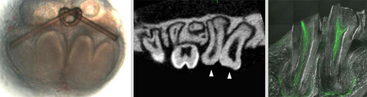
Left: Tooth germ split into two after ligation. Middle: Micro-CT of split tooth germ 50 days after transplantation into oral cavity of mouse.
Right: Nerve fibers in the tooth pulp and periodontal ligament of split tooth.
The tooth germ is derived from the embryonic ectoderm through epithelial-mesenchymal interactions. The team first extracted molar tooth germ from the jaw of an ED 14.5 mouse, when the tooth germ is generated, and tied a knot (ligation) around the tooth germ using a thin nylon thread, specifically through the signaling center in an attempt to split the tooth germ. After culturing the ligated tooth germ for six days, histological stainings revealed the formation of two tooth germs, each surrounded by epithelial tissue. The ligated tooth germ were then transplanted into the mouse subrenal capsule, and after 30 days of maturation, it gave rise to two teeth that contained hard tissues such as enamel and dentin, which was surrounded by periodontal ligament and alveolar bone, as seen in natural teeth. The split teeth however were half the size of natural molar teeth, and also had half the number of dental cusps on the crown of the tooth.
Next, the team carefully analyzed the germ splitting process using time-lapse imaging, and found that after ligation, the tooth germ signaling center, enamel knot, appeared in each split germ. Epithelial tissues surrounding each split germ continued to proliferate, eventually invaginating along the ligated surface resulting in two separate germs. Gene expression analyses for genes important in early tooth development showed split germs displayed similar expression patterns as those in natural tooth germ.
Past studies have shown that tooth morphology is regulated by reaction-diffusion waves of gene expression involving an activator, Lef1, and an inhibitor, Ectodin. Analyses of the expression patterns of these two genes were performed in natural and split tooth germs. In natural tooth germ, Lef1 was expressed in the enamel knot and neighboring mesenchyme, and Ectodin was expressed around the region of Lef1 expression. The split tooth germ showed smaller yet comparable expression patterns within each germ. Thus, ligation of the tooth germ leads to re-regionalization of the tooth-forming field in each split germ through reaction-diffusion waves of gene expression.
The group then implanted the split tooth germ into the jaw bone of the mouse to test whether teeth would develop and erupt similar to natural tooth. The transplanted germs became engrafted into the surrounding tissue, and two months post-transplantation, micro-CT analysis revealed that the newly erupted teeth were aligned and in contact with the teeth on the opposing jaw. When orthodontic methods similar to braces were applied to the split teeth, the bone supporting the teeth underwent remodeling to accommodate teeth movement, indicating the presence of a functional periodontal ligament. Nerve fibers were also detected in the tooth pulp and periodontal ligament of the split teeth, as seen in natural teeth. Analyses of response to noxious stimuli, such as orthodontic force or pulp exposure, revealed both engrafted and natural teeth showed comparable responses, indicating that split teeth were properly connected to the central nervous system.
“Not only does tooth loss affect physiological tooth functions such as teeth alignment, chewing and speech, it can also affect overall health,” explains Tsuji. “In the future, our method could be used as a new form of regenerative medicine therapy for patients with tooth loss by generating multiple tooth germs from the patient’s own tooth germs and later implanting them.”
| Link to article |
|---|
![CDB [RIKEN CENTER FOR DEVELOPMENTAL BIOLOGY]](http://www.cdb.riken.jp/en/wp-content/themes/cdb_en/images/common/fLogo2.png)
2-2-3 Minatojima-minamimachi, Chuou-ku, Kobe 650-0047, Japan
TEL : +81-78-306-0111
FAX : +81-78-306-0101
E-mail : cdb[at]cdb.riken.jp

