
News and Announcements from the CDB
In utero electroporation is a technique that is widely used in the study of brain development to introduce DNA or RNA into the brain of the developing embryo inside the uterus by applying electrical pulses. This method can be used to label cells by inducing fluorescent protein expression vectors, or to modify gene function by overexpression, mis-expression or knocking down expression of a specific gene and examining phenotypes at tissue level. It remained difficult, however, to modify the gene itself using in utero electroporation. If gene knock-in was made possible via in utero electroporation, it would be possible to alter the genome of a specific cell population in the brain during development, facilitating labeling of cells, lineage tracing, or analyses of the localization and dynamics of a protein at the cellular level in vivo.
Research scientist Yuji Tsunekawa, student trainee Raymond Terhune and others in the Laboratory for Cell Asymmetry (Fumio Matsuzaki, Team Leader) have now developed a new tool for introducing genes into the developing mammalian brain by combining the use of the CRISPR/Cas9 system and in utero electroporation. In a paper published in Development, they demonstrate the high efficiency of their gene knock-in method to insert transgenes into a target site in neural progenitors in the embryonic mouse brain. Furthermore, the team refined their protocol to insert two differently fluorescent markers into a target gene in each homologous chromosome, which enabled them to visualize by color the cells with homozygous knocked-in alleles.
The success of a gene knock-in protocol is dependent on the efficacy of gene targeting events, which is usually very low. Invention of the CRISPR/Cas9 system has enabled the genome to be edited with ease and high precision. Tsunekawa and his collaborators have been working to develop CRISPR/Cas9-based gene-editing techniques that can efficiently modify the genome, and have more universal applications, one of which culminated in the development of HITI, which can efficiently knock-in genes in differentiated cells in vivo (see Science News: January 11, 2017). Concurrently with the development of HITI, the team tried developing a simple gene knock-in method targeting neural progenitor cells in embryonic brain in utero by combining the CRISPR/Cas9 method with in utero electroporation.
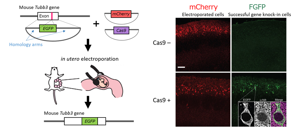
CRISPR/Cas9 system-based genome-editing technology exploits the two intrinsic DNA repair pathways of cells: homology-directed repair (HDR) and non-homologous end joining (NHEJ). Whereas the HITI approach used the NHEJ pathway to insert genes into both proliferating and non-proliferating cells, to specifically target proliferating neural progenitors in the brain, the team elected to use the HDR pathway, which functions only in actively dividing cells but is more accurate and efficient. They first designed the vector such that the EGFP sequence to be inserted was flanked by homology arms and tested their method to knock-in EGFP (green) fluorescent marker targeted to the Tubb3 gene, a neural marker. They found EGFP was knocked-in in approximately 20% of the electroporated cells. Proving that their concept did work, the team then determined the best homology arm lengths for efficient gene insertion and optimizing the vector template to minimize leaky expression.
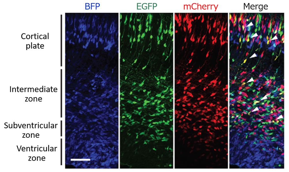
Genes knocked-in to the cell are inserted at random into one target loci of homologous chromosomes, thus resulting in cells carrying gene insertions in either one of the two loci (heterozygous) or in both loci (homozygous) of homologous chromosomes. The team wondered whether it was possible to distinguish between heterozygous and homozygous targeted cells, and designed a protocol in which two targeting vectors, each with a different colored fluorescent marker, were electroporated simultaneously in utero into the mouse brain. The theory was that cells in which both loci had been knocked-in would fluoresce both colored signals to produce a different colored signal, thus indicating a cell carried homozygous knocked-in alleles. When the team tested their protocol targeting Tubb3 gene with EGFP (green) and mCherry (red), they found cells expressing both EGFP and mCherry produced a yellow signal, indicating that donor sequence had been knocked-in to both Tubb3 gene loci of those cells (approximately 5 % of the electroporated cells).
“Efficient observation of simultaneous fluorescence of two different colored signals is made possible by the CRISPR/Cas9 system’s efficiency for introducing transgenes. Using this method, we can now identify cells with homozygous knocked-in alleles, and also trace cell lineages with live-imaging,” explains Matsuzaki. “We also showed that our new protocol can be used in ferret models, demonstrating that this can be used for non-rodent animals for which generating genetically modified animals is difficult, and spur research advances in neurosciences.“
| Link to article | |
|---|---|
| Related link |
![CDB [RIKEN CENTER FOR DEVELOPMENTAL BIOLOGY]](http://www.cdb.riken.jp/en/wp-content/themes/cdb_en/images/common/fLogo2.png)
2-2-3 Minatojima-minamimachi, Chuou-ku, Kobe 650-0047, Japan
TEL : +81-78-306-0111
FAX : +81-78-306-0101
E-mail : cdb[at]cdb.riken.jp

