
News and Announcements from the CDB
Cell division is a finely tuned process involving the partitioning of cytoplasm and the segregation of chromosomes into two daughter cells. Normally, paired chromosomes align along the spindle equator formed by microtubules, which then pulls the paired chromosomes to opposing cell poles. In somatic cells, mistakes in chromosome segregation are rarely observed thanks to a stringent checkpoint mechanism that ensures all chromosomes are correctly aligned before segregation begins. In contrast, oocytes, the female germ cells involved in reproduction that undergo meiotic cell division, display a surprisingly high rate of chromosome segregation errors (approximately 10–30%), which can lead to miscarriages or congenital diseases. The complete causal mechanisms underlying the high frequency of errors in chromosome segregation seen in oocyte meiosis remain largely unknown.
Now, a new study led by research scientist Hirohisa Kyogoku in the CDB’s Laboratory for Chromosome Segregation (Tomoya Kitajima, Team Leader) examined the effects of cytoplasmic size on chromosome segregation in oocytes during the first meiotic division by manipulating the cytoplasmic volume of mouse oocytes. In their work published in Developmental Cell, the team demonstrated that oocytes with larger cytoplasm are more prone to display broadly dispersed spindles and a less rigorous spindle checkpoint before progression to anaphase, the cell-cycle phase when chromosomes begin segregating, which contributes to increased frequency of errors in chromosome segregation and aneuploidy.
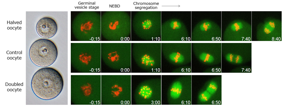
The oocyte’s large cytoplasm is one feature that makes it distinct from other somatic cells, and the team hypothesized that this large cytoplasmic size was somehow linked to the higher frequency of chromosome segregation errors observed during meiosis. To test this, they made oocytes containing half the amount of cytoplasm (i.e. halved oocytes) and those containing double the amount of cytoplasm (i.e. doubled oocytes) found in normal oocytes, using micromanipulation. The halved oocytes were created by aspirating half of the cytoplasm from an oocyte using a micropipette, while the doubled oocytes were created by fusing a normal oocyte with an enucleated oocyte. They also confirmed that these artificially created oocytes could be induced to enter the first meiotic division at rates comparable to control oocytes.
Next, the team used live-cell imaging to track the movements of the chromosomes and the meiotic spindle within the oocytes undergoing meiosis, and carried out quantitative analyses of their three dimensionally reconstructed data. They first noticed a correlation between the sizes of the oocyte cytoplasm and that of the spindle—as cytoplasmic size increased, so did the relative spindle size. In addition, when they examined the dynamics of the oocyte’s microtubule-organizing centers (MTOCs), which are not anchored by centrosomes as seen in somatic cells, the team observed that those with larger cytoplasm displayed less dense and more broadly dispersed MTOCs. The MTOCs in oocytes initially appear as a spherical mass of microtubules, which gradually splits to relocate to two opposite poles creating spindle bipolarization. The doubled oocytes showed spindle formations at both poles that covered a larger area than those seen in control and halved oocytes and that were also more prone to separate into smaller units, contributing to errors in chromosome alignment along the spindle equator. Oocytes with larger cytoplasm were also unable to align chromosomes at the spindle equator as efficiently as compared to those with relatively smaller cytoplasm, requiring more time for proper alignment.
Normally, if chromosomes fail to align properly along the spindle equator, the spindle checkpoint mechanism would be triggered, blocking entry into anaphase until the errors are fixed. In oocytes, this checkpoint mechanism appears to be less rigorous, with some entering anaphase despite mistakes in chromosome alignment. Molecular factors involved in the checkpoint mechanism are produced in the nucleus and are released into the cytoplasm when the nuclear envelope breaks down at the entry of meiosis. Kyogoku and Kitajima found that these checkpoint factors become diluted within the cytoplasm as cytoplasmic size increases which results in a weakened checkpoint system that permits chromosomes to segregate prematurely before they are all properly aligned. The checkpoint mechanism was more stringent in the halved oocytes, halting anaphase entry when even one chromosome was improperly aligned, whereas anaphase entry was seen many doubled oocytes despite errors in chromosome alignment leading to errors in chromosome segregation.
“We were able to demonstrate experimentally that the large cytoplasm is one factor contributing to the high frequency of chromosomal segregation errors seen in oocytes. As the large cytoplasm is considered to be crucial for supporting early embryonic development, our findings suggest that there is trade-off between accuracy of meiosis and post-developmental competence,” explains Kitajima. “There are many other causes of chromosome segregation errors in oocytes, such as maternal aging. By understanding the different causes of chromosome segregation errors, we hope to be able to impart insights for developing strategies to prevent or rescue chromosome segregation errors.”
| Link to article |
Large Cytoplasm Is Linked to the Error-Prone Nature of Oocytes |
|---|---|
| Related link |
![CDB [RIKEN CENTER FOR DEVELOPMENTAL BIOLOGY]](http://www.cdb.riken.jp/en/wp-content/themes/cdb_en/images/common/fLogo2.png)
2-2-3 Minatojima-minamimachi, Chuou-ku, Kobe 650-0047, Japan
TEL : +81-78-306-0111
FAX : +81-78-306-0101
E-mail : cdb[at]cdb.riken.jp

