
News and Announcements from the CDB
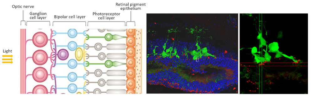
The retina is a layered neural structure at the back of the eye that plays an important role in sensing and translating light information entering the eye into neural signals sent to the brain. Light is sensed by photoreceptors (rods and cones) in the outer nuclear layer, the outermost layer of the neural retina, and this information is then transmitted in turn through the bipolar cell layer, ganglion cell layer, and finally to the optic nerve that will send neural signals to the brain to be interpreted into what we “see.” Disruption to any segment of this neural transmission can affect vision. Retinitis pigmentosa is a group of genetic eye disorders characterized by progressive degeneration of retinal photoreceptors resulting in severe visual impairment, and there is currently no effective treatment to restore visual function in affected patients. Some groups have reported that transplantation of retinal tissue or stem cell-derived photoreceptors into the eye shows slight restoration of visual function, but it remained unclear whether transplanted tissues or photoreceptors actually integrated with the surrounding host environment.
A new study led by Deputy Project Leader Michiko Mandai of the Laboratory for Retinal Regeneration (Masayo Takahashi, Project Leader) has demonstrated that following transplantation of induced pluripotent stem cell (iPSC)-derived retinal tissue into end-stage retinal degeneration (rd1) model mice, the photoreceptors in the transplanted tissue can form functional synapses with synaptic partners in the host eye using cell labeling, behavioral analyses and electrophysiological recordings. Their work was published in the journal Stem Cell Reports.
A series of studies by the former CDB Laboratory for Organogenesis and Neurogenesis led by the late Yoshiki Sasai established a cell culture method for directing embryonic stem cells (ESCs) to self-organize into 3D tissue structures and also reported the successful generation of 3D-retinal tissue from mouse and later human embryonic stem cells (ESCs) (Science News: April 7, 2011; June 14, 2012). Mandai and her team expanded on this work to examine feasibility of using this stem cell-derived retinal tissue for transplantation to restore vision in retinal degenerative diseases. They reported that mouse ESC/iPSC-derived retinal tissue could mature and survive when transplanted into mouse eye (Science News: April 25, 2014), and observed similar results transplanting human ESC-derived retinal tissue in monkey models (Science News: January 29, 2016). Although the retinal grafts appeared to integrate with surrounding host retina in both studies, they could not ascertain whether the graft had fully integrated with the host to sense light and transmit neural signals.
The group first examined the synaptic connections between the photoreceptors in transplanted tissue and the surrounding host cells, specifically with bipolar cells. They generated and transplanted iPSC-derived retina expressing red fluorescent markers at the synaptic terminal ends of photoreceptors into an rd1 mouse line that expressed green fluorescent markers in dendrites of bipolar cells. They were able to visually confirm that the red photoreceptor synaptic terminal ends of their transplanted retinal tissue were in contact with green host bipolar cell dendrites.
Next, they used a behavioral learning experiment, called shuttle avoidance system (SAS), on rd1 mice to determine whether these mice could in fact detect light following transplantation of iPSC-derived retina. In SAS, mice can be trained to associate stimulus, such as light, with electrical shock and will move to avoid getting shocked when they sense light. Thus, if the iPSC-derived retina had formed functional synapses with host cells, the transplant recipient rd1 mice should be able to be trained to associate light stimulus with an electrical shock and show avoidance behavior if light is detected. Their analyses revealed that some transplant recipient rd1 mice displayed avoidance responses, while rd1 mice that did not receive transplants moved at random regardless of light stimulus.
The team also extracted the whole retina of rd1 mice post-transplantation to examine their ability to transmit electrophysiological signals using a microelectrode array (MEA) system. The extracted whole retina tissues were laid flat on microchips and analyzed for their responses to light signals. The team found that the retinal graft areas responded to light signals similar as seen in normal retina, and that graft-derived photoreceptors transmitted excitatory signals via bipolar cells to host retinal ganglion cells.
“Presently, we can only transplant tissue sizes equivalent to less than 5% of the whole retina with our method. If we can improve our technique to allow transplantation of larger tissues, this may lead to a marked improvement in vision,” says lead author Mandai. “Our study demonstrates a proof-of-concept for considering clinical transplantation of iPSC-derived retinal tissues into patients with retinal degeneration. Aside from improving transplant techniques and testing whether human iPSC-derived retina can restore vision in blind mice, we also need to assess the safety of the transplant procedure and of the derived retinal tissue itself before we can move to human clinical studies.”
![CDB [RIKEN CENTER FOR DEVELOPMENTAL BIOLOGY]](http://www.cdb.riken.jp/en/wp-content/themes/cdb_en/images/common/fLogo2.png)
2-2-3 Minatojima-minamimachi, Chuou-ku, Kobe 650-0047, Japan
TEL : +81-78-306-0111
FAX : +81-78-306-0101
E-mail : cdb[at]cdb.riken.jp

