
News and Announcements from the CDB
Cells of an animal’s immune system are capable of differentiating between self and non-self cells by recognizing proteins expressed on the cell surface called, major histocompatibility complexes (MHC). In humans, these cell surface proteins are called human leukocyte antigens (HLA). When immune cells encounter MHCs they do not recognize, an immune response is triggered to rid the body of the foreign entity. This mechanism is important for protecting the body from pathogens, but can be a hindrance when considering cell or organ transplantation from one individual to another.
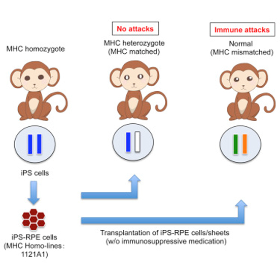
Now a research team led by Sunao Sugita, deputy project leader of the Laboratory for Retinal Regeneration (Masayo Takahashi, Project Leader) and other colleagues has demonstrated experimentally that immune responses can be avoided when retinal pigment epithelial (RPE) cells generated from induced pluripotent cells (iPSCs) derived from a MHC/HLA homozygous donor are matched with MHC/HLA of recipient. The research team developed an in vivo experimental method in a primate model, in which they transplanted RPE cells derived from iPSCs from a MHC homozygote monkey into a different monkey, and an in vitro experimental system using RPE cells derived from human iPSCs and co-cultured them with human immune cells, including T cells. Their study was published in two separate papers in Stem Cell Reports.
The laboratory has been working to establish treatments for retinal diseases using iPSC-derived cells. They developed a method to generate RPE cells from human iPSCs, and in 2014, performed the first autologous RPE transplantation into a human patient with age-related macular degeneration (AMD), a disease that damages the RPE cell layer of the retina (see Science news: Sep. 15, 2014). As the original cell source for autologous transplantation is the patients themselves—iPSCs derived from patient’s own cells are differentiated into RPE cells, and later transplanted back into the patient—the potential for rejection was considered minimal. However, as this method is costly and time-consuming, the group has been considering allogeneic transplantation (iPSC-derived cells generated from one person transplanted into another person) as an approach to overcome these issues. While allogeneic transplantation raises concerns for risk of immune rejection and need for use of immunosuppressant drugs, in theory, matching MHC/HLA of graft and recipient should reduce potential for immune rejection, similar to organ transplantation, but this had not been tested before for iPSC-derived cells.
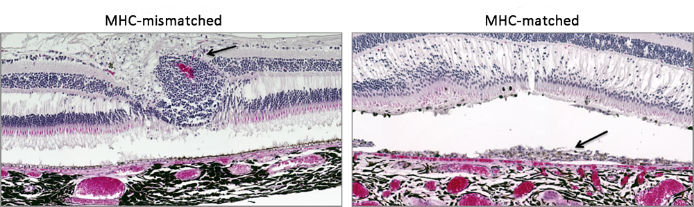
The team generated RPE cells and sheets from monkey iPSC lines with an MHC homozygous profile, and then transplanted these RPE cells or sheets into the sub-retinal space of the eyes of MHC-matched or mismatched monkeys. In MHC-mismatched monkeys, the team observed retinal tissue damage and signs of immune rejection, such as infiltration of inflammatory cells into the subretinal space, following transplantation. On the other hand, no signs of immune rejection were observed in recipients of MHC-matched grafts, and the sheet grafts survived for at least six months post-transplantation without the need to use immunosuppressant drugs.
Sugita and his colleagues also examined immunogenicity using an in vitro experimental system with human iPSC-derived RPE cells. They generated iPSC lines from HLA homozygous donors, and then differentiated them into RPE cells, which were then co-cultured with lymphocytes isolated from a different donor. The researchers found that when HLA types were mismatched, there was an increase in inflammatory cell numbers as well as a rise in levels of IFNγ inflammatory cytokine, similar to what is observed in early stages of an immune response. In contrast, the matching of at least three HLA gene loci (HLA-A, HLA-B, and HLA-DRBI) between iPSC-derived RPE cells and the donor immune cells was sufficient for preventing the trigger of an immune response. The HLA matching of these three loci have been known to be critical when considering organ transplantation or bone marrow transplants, and the present study demonstrated for the first time that the same is true for allogeneic transplantation of iPSC-derived RPE cells.
The laboratory announced the start of a new clinical research study in June this year (see Science news: Jun. 8, 2016), which includes plans to use the iPSC bank established at the Center for iPS Cell Research and Application (CiRA) at Kyoto University to derive RPE cells for allogeneic transplantations.
“The potential risks posed to the whole body in using immunosuppressant drugs is a high price to pay when considering the size of the eye. But, our current study shows that these drugs may not be needed if at least three HLA loci are matched,” explains Sugita. “While RPE cells were the focus of our studies, the in vivo and in vitro experimental systems that we developed may be adaptable and prove useful for other groups working towards iPSC-based clinical applications in other tissues or organs.”
| Link to article | |
|---|---|
| Related links | |
![CDB [RIKEN CENTER FOR DEVELOPMENTAL BIOLOGY]](http://www.cdb.riken.jp/en/wp-content/themes/cdb_en/images/common/fLogo2.png)
2-2-3 Minatojima-minamimachi, Chuou-ku, Kobe 650-0047, Japan
TEL : +81-78-306-0111
FAX : +81-78-306-0101
E-mail : cdb[at]cdb.riken.jp

